
Case Quiz December 2025
- Patient: Canine, Prague Ratter, 2 years, F
- Presenting Signs: Acute onset and one week duration of cough, tachypnea, mixed dyspnea and fever.
Case of the month: December 2025
Patient:
Canine, Prague Ratter, 2 years, F
History:
Acute onset and one week duration of cough, tachypnea, mixed dyspnea and fever.
Findings:
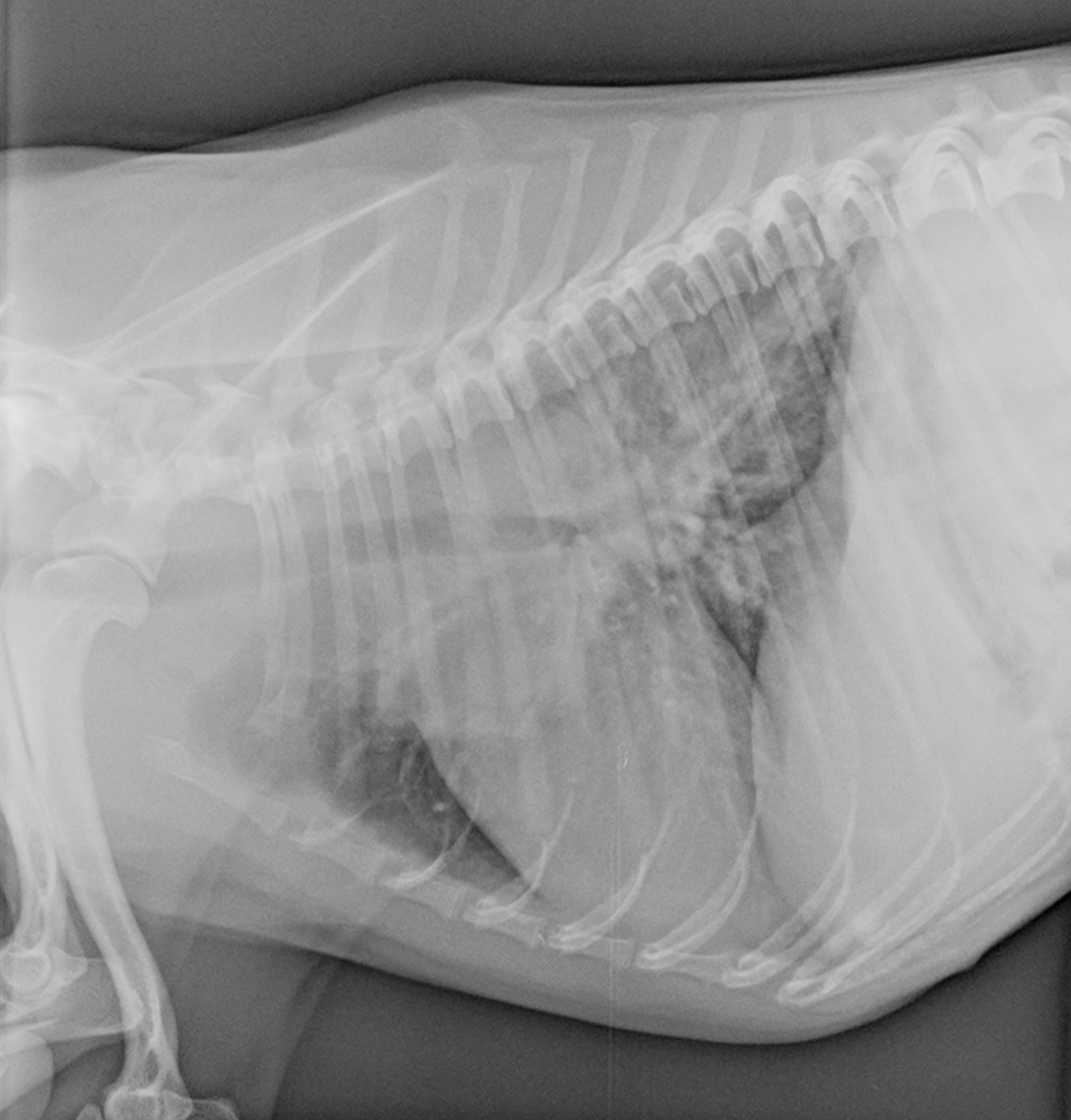
Two sets of radiographs, taken 5 days apart, are available for review. Each contains three views of the thorax (left and right lateral, and ventrodorsal). Images are of good quality, with only slight patient rotation on the lateral views in the first set.
A diffuse, unstructured interstitial lung pattern with peribronchial cuffing is visible on both sets of radiographs. Worst on the second set. A soft-tissue opacity is visible in the cranial right lung lobe. It is better visualised in the second set of radiographs, where the lobar sign is present.
The trachea, mediastinum, pleural spaces, heart, pulmonary vessels, thoracic walls, and diaphragm are unremarkable.
Differential diagnoses:
- granulomatous pneumonia
- primary and metastatic pulmonary neoplasia
- immune-mediated pulmonary disease
- pulmonary thromboembolism
- pulmonary haemorrhage
Conclusion/Diagnosis:
A CT examination, bronchoscopy and fine-needle biopsy were scheduled.
The CT scan confirmed a soft-tissue attenuating mass with a mild heterogeneous contrast enhancement within the right cranial lung lobe with well-defined margins. Mild compression of the left cranial lung lobe and left-sided mediastinal shift. Multifocal peribronchial ground glass opacities within the dorsal third of multiple lung lobes were identified.
The samples obtained via bronchoscopy and US-guided fine-needle biopsies were submitted for cytological examination. The results suggested an eosinophilic granuloma.
Outcome:
There was a clinical improvement after two days of conservative treatment. After two months, the pulmonary mass was no longer visible on radiographs or ultrasound images.
Discussion:
Details regarding the laboratory findings and therapy are available in the published paper cited below.
The Discussion in the paper contextualises the results, highlighting that pulmonary eosinophilic granuloma (PEG) is currently considered a rare interstitial lung disease (ILD) with a guarded prognosis due to frequent relapses. It encourages readers to review the established diagnostic criteria met in this case, including imaging and elevated eosinophil counts in the BAL fluid. Furthermore, the discussion delves into the potential connection between PEG and a systemic immune process, hypothesising that the patient’s subsequent anaphylactoid reaction to routine vaccination suggests a systemic manifestation of allergic disease. The authors explain the rationale for combined immunosuppressive therapy, which enabled minimised steroid dosage and allowed the dog to remain disease-free for almost three years, challenging previous guarded prognoses for
PEG and supporting documentation of prolonged survival. This comprehensive analysis provides valuable insights into the diagnosis, complex pathophysiology, and successful long-term management of canine PEG.
Further reading:
Agudelo CF, Stehlik L, Filipejova Z, Koskova B, Sterbova M, Crha M. Pulmonary
eosinophilic granulomatosis in a dog. Vet Med (Praha). 2022 Jan 18;67(3):150-155.
Doi: 10.17221/136/2020-VETMED
For full case clickhere.
Smarter Teleradiology. Faster Diagnosis. Better Results Join the Veheri Community!
Start today: Book your DEMO