
CT of adrenal masses in dogs
What contrast CT shows in a dog with an adrenal mass, from caval invasion and adhesions to tumour type, and where its accuracy runs out before adrenalectomy.

Referenced guides for veterinarians on when CT or MRI answers the clinical question, by species and body region.
What contrast CT shows in a dog with an adrenal mass, from caval invasion and adhesions to tumour type, and where its accuracy runs out before adrenalectomy.

What brain CT can and cannot detect in dogs when MRI is not available, for masses, stroke, inflammation and head trauma, and when MRI or CSF is still needed.
When CT is worth it for medial coronoid disease in dogs, what it finds that radiographs miss, and where arthroscopy still gives the better answer.
When abdominal CT adds to radiographs and ultrasound in vomiting dogs with suspected obstruction, foreign body or perforation, and what CT can and cannot show.
What contrast CT adds for a dog with a renal mass or a dilated renal pelvis, which features point to carcinoma or lymphoma, and what CT cannot decide.
How multiphase CT characterises a focal liver mass in dogs, how reliable its benign versus malignant signs are, and what it adds to surgical planning.
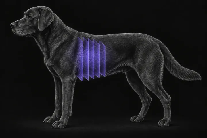
How thoracic CT compares with radiographs for pulmonary metastasis screening in dogs, which tumours most often spread to the lung, and how to read a nodule.
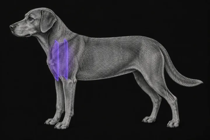
How CT helps separate thymic epithelial tumours from lymphoma in dogs with a cranial mediastinal mass, and what it shows about invasion and surgical approach.
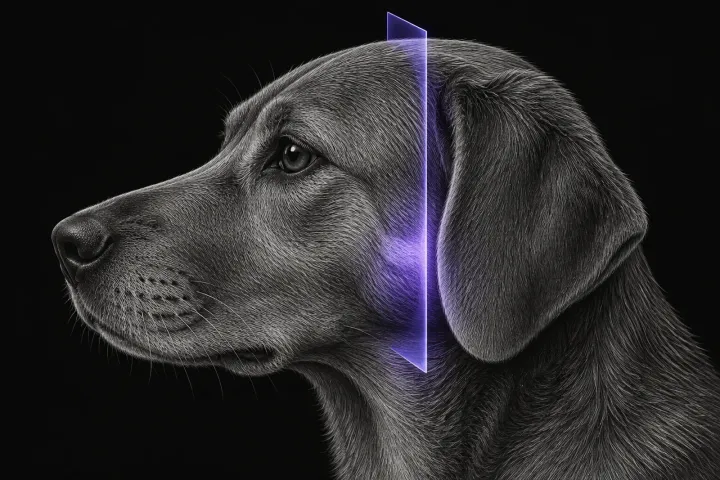
How CT detects otitis media in dogs, how it compares with otoscopy, ultrasound and MRI, and which CT signs point towards cholesteatoma or neoplasia.
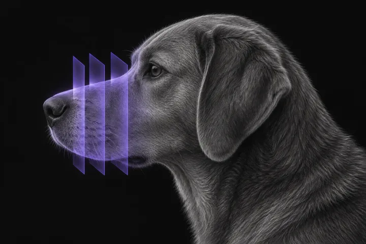
How CT separates nasal tumours, fungal rhinitis, foreign bodies and inflammatory rhinitis in dogs with chronic nasal discharge, and where rhinoscopy and biopsy are still needed.
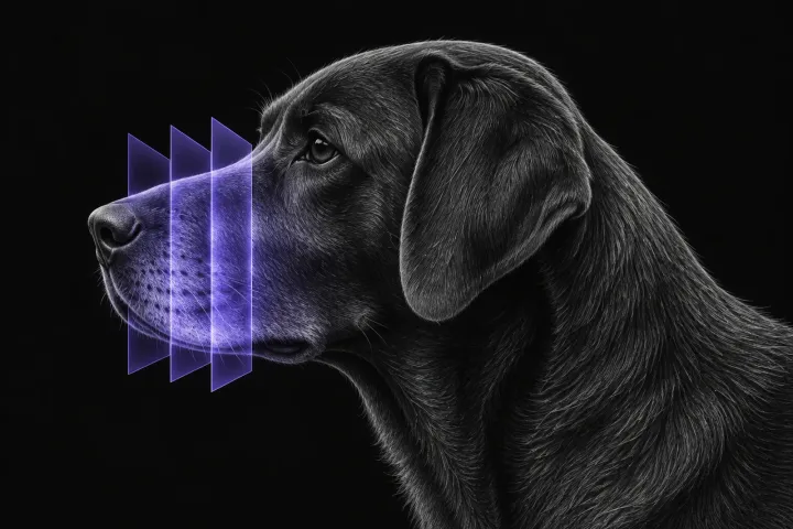
How CT stages canine oral tumours, from bone invasion to the mandibular and medial retropharyngeal nodes, and why node sampling is still needed.
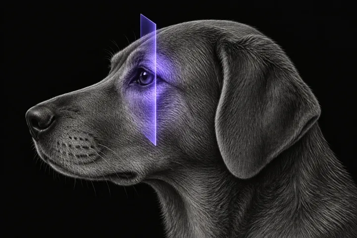
How CT helps separate orbital neoplasia from cellulitis and other inflammation in dogs with exophthalmos, where MRI adds detail, and how CT guides sampling.
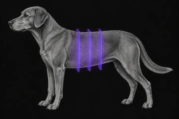
How multiphase CT localises a suspected insulinoma in dogs, why arterial timing matters, how well it finds metastases, and where surgery has the last word.
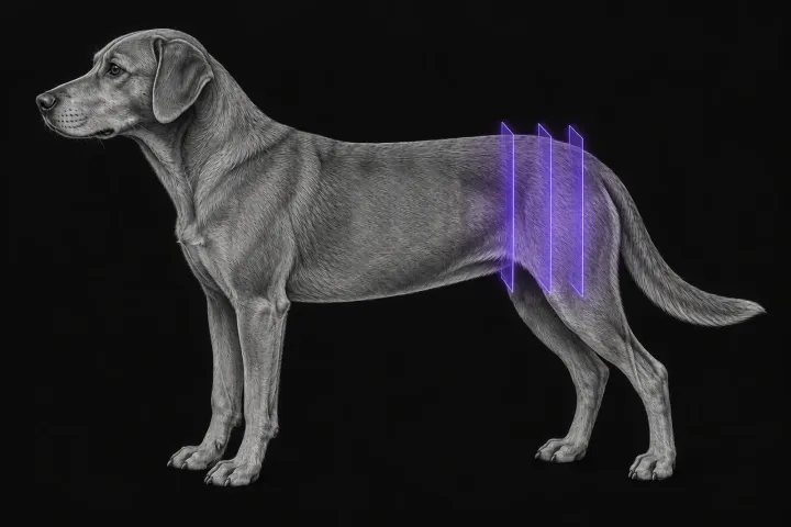
When CT adds to radiographs after canine pelvic trauma, which fractures it finds more reliably, and how to use 2D and 3D images for surgical planning.
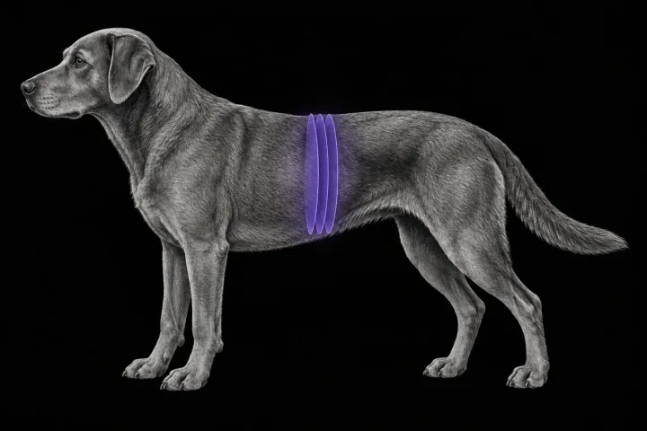
How CT angiography compares with ultrasound for congenital portosystemic shunts in dogs, how shunts are classified, and what the surgeon needs from the report.
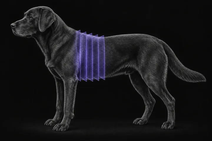
When to request CT pulmonary angiography for suspected pulmonary thromboembolism in dogs, what a positive scan looks like, and why technique decides the answer.
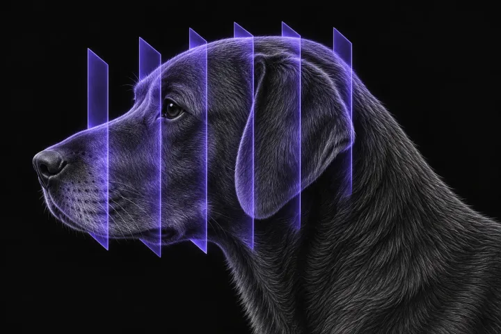
What head CT shows after trauma in dogs, which skull fracture patterns matter, and how far CT findings can support a prognosis alongside the neurological exam.
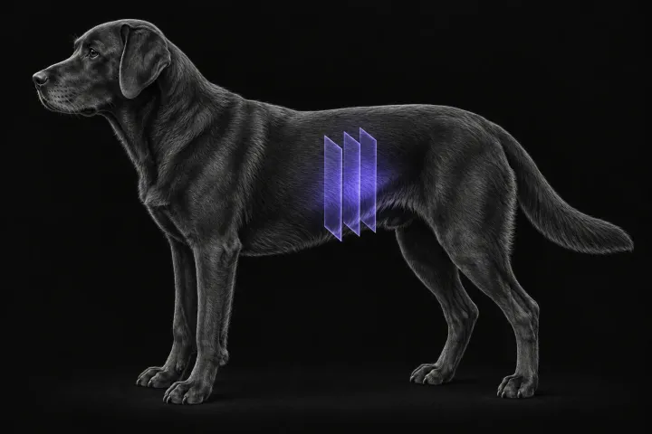
What contrast CT can and cannot tell you about a canine splenic mass, why benign and malignant lesions overlap, and where CT adds most to staging.
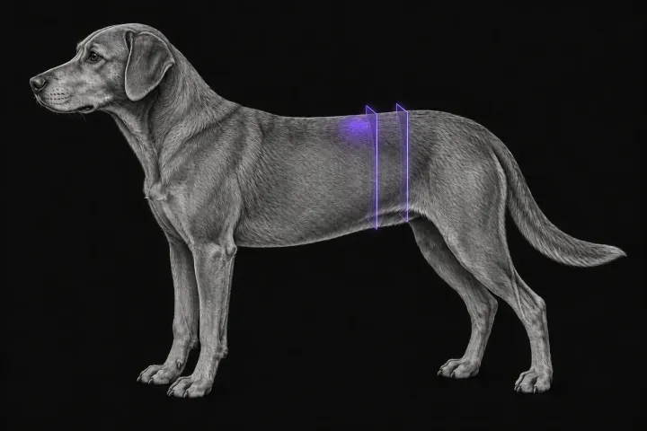
How CT compares with abdominal ultrasound for staging the iliosacral lymph nodes in dogs with anal sac adenocarcinoma, and what the report should state.
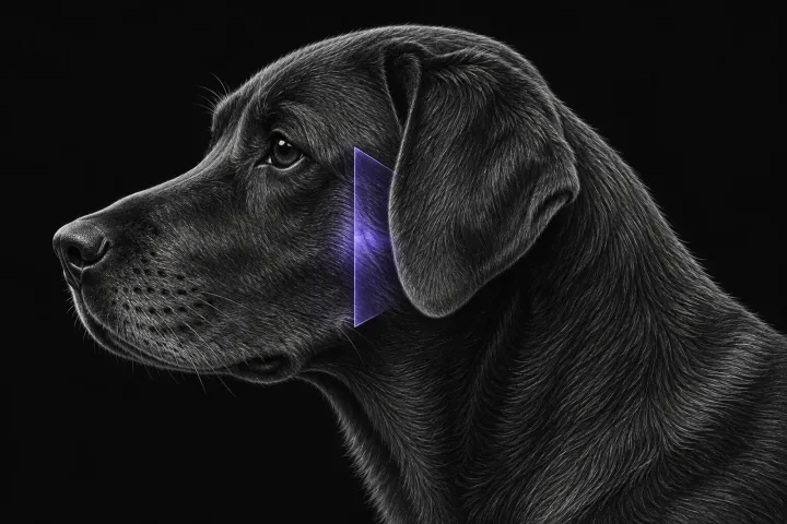
When CT of the canine temporomandibular joint is indicated, what it shows in fracture, ankylosis, dysplasia and osteoarthritis, and where it falls short.
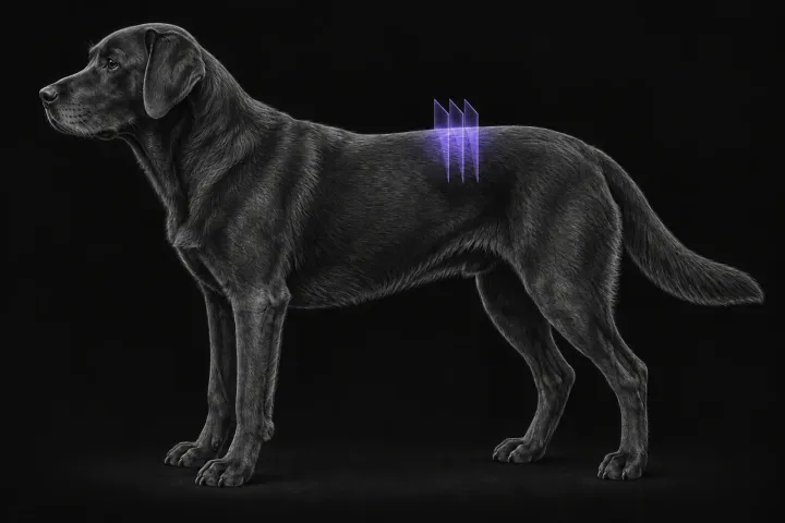
When non-contrast CT is enough for thoracolumbar disc extrusion in chondrodystrophic dogs, and which dogs need CT-myelography or MRI instead.
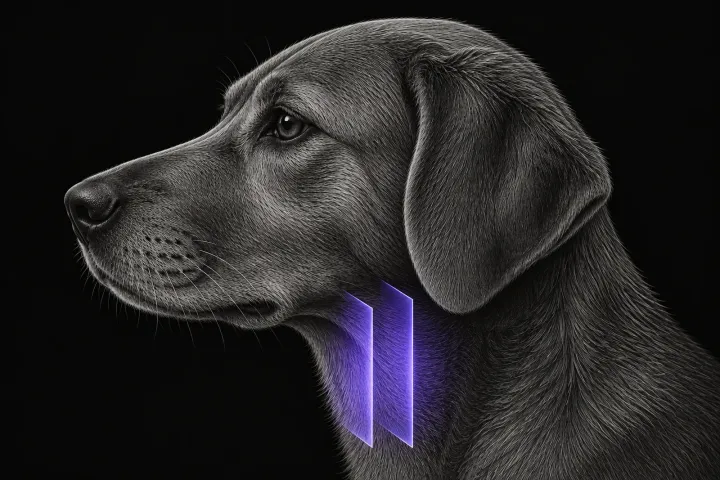
What CT adds to palpation and ultrasound in dogs with a thyroid mass, how well it shows invasion, and what it means for thyroidectomy and staging.
How CT excretory urography compares with ultrasound and cystoscopy for ectopic ureters in dogs, and what the report must state before laser ablation or surgery.
What contrast CT shows in feline injection-site sarcoma, how it changes surgical and radiotherapy planning, and where imaging cannot replace histology.
What thoracic CT adds over radiographs in cats with lung tumours, bronchial or interstitial disease, how anaesthesia affects the lungs, and where CT stops.
What CT adds for a cat with a cranial mediastinal mass, how far it separates thymoma, lymphoma and cysts, and why cytology or histology still decides.
When to image the feline tympanic bulla, how CT compares with radiography, ultrasound and MRI, and what CT shows in otitis media, polyps and middle ear neoplasia.
Which CT features separate nasal lymphoma, carcinoma, fungal and chronic rhinitis in cats, why the retropharyngeal nodes matter, and why biopsy is still needed.
What CT shows in nasopharyngeal polyps, stenosis, lymphoma and cryptococcosis in cats with stertor, and when endoscopy and biopsy are still needed.
How CT stages oral squamous cell carcinoma in cats, what bone lysis and orbital extension look like, how often nodes are involved, and where CT falls short.
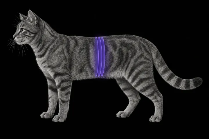
How CT angiography maps portosystemic shunts in cats, how it compares with ultrasound and portovenography, and which feline anatomy the report must cover.
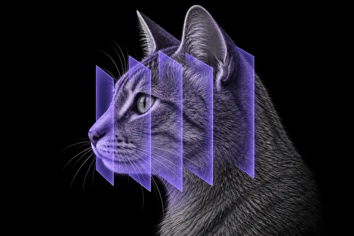
Which skull, jaw and temporomandibular joint fractures CT finds in cats after head trauma, why radiographs miss them, and which findings change treatment.
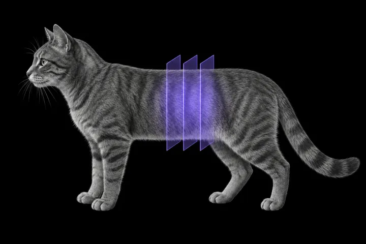
How non-enhanced CT compares with ultrasound for ureteroliths in cats, why calculus count and position matter, and what CT cannot show.
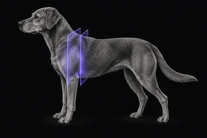
How MRI finds peripheral nerve sheath tumours of the canine brachial plexus, what the radiologist looks for, and what imaging can say about grade and extent.
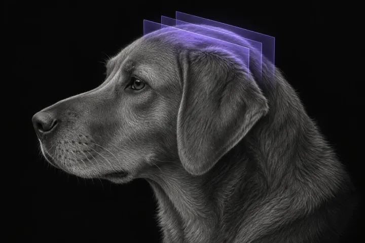
Which dogs with seizures need brain MRI, what the evidence says about diagnostic yield, what the radiologist looks for and what your report will include.
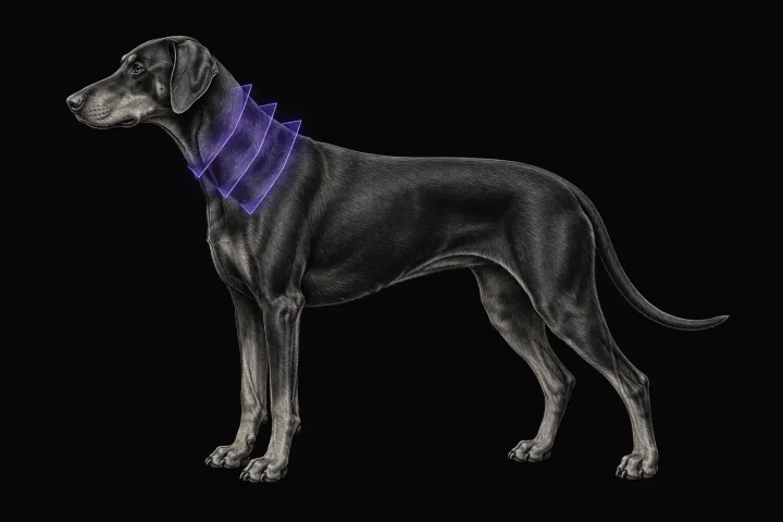
How MRI characterises cervical spondylomyelopathy in dogs, what traction and flexion-extension studies add, and why compression alone does not prove the cause.
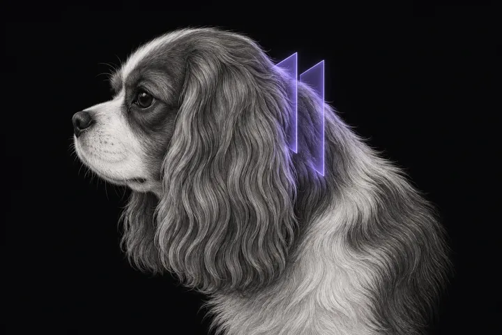
What MRI of the canine craniocervical junction should include for Chiari-like malformation and syringomyelia, and how to read a syrinx against the signs.
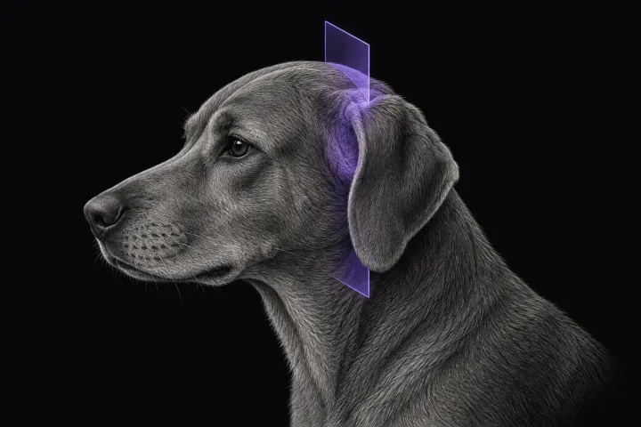
How reliably the neurological exam separates central from peripheral vestibular disease in dogs, and what MRI shows in otitis interna and idiopathic cases.
What MRI shows in degenerative lumbosacral stenosis in dogs, which findings relate to clinical signs, and why changes in normal dogs need context.
What MRI and CT show in canine masticatory muscle myositis, how imaging guides muscle biopsy, and which other causes of jaw muscle atrophy it must exclude.
When MRI or CT best answers canine orbital and optic nerve questions, which signs point to tumour or inflammation, and when sampling is still needed.
When pituitary MRI helps in dogs with pituitary-dependent hypercortisolism, what the P/B ratio means, and how tumour size shapes surgery and radiotherapy.
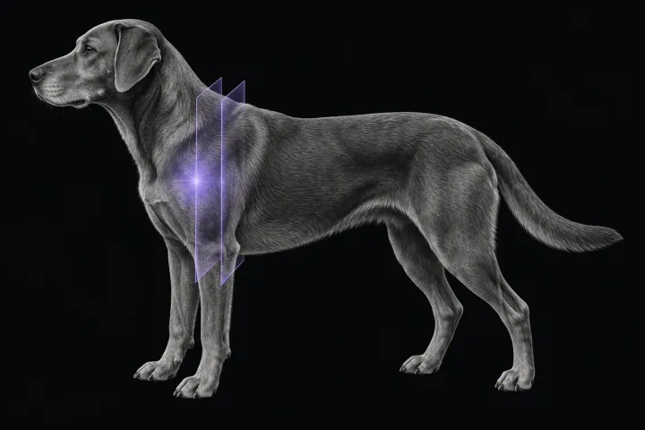
What MRI shows in canine biceps and supraspinatus tendinopathy, how it compares with CT arthrography, and why a bright tendon alone is not disease.
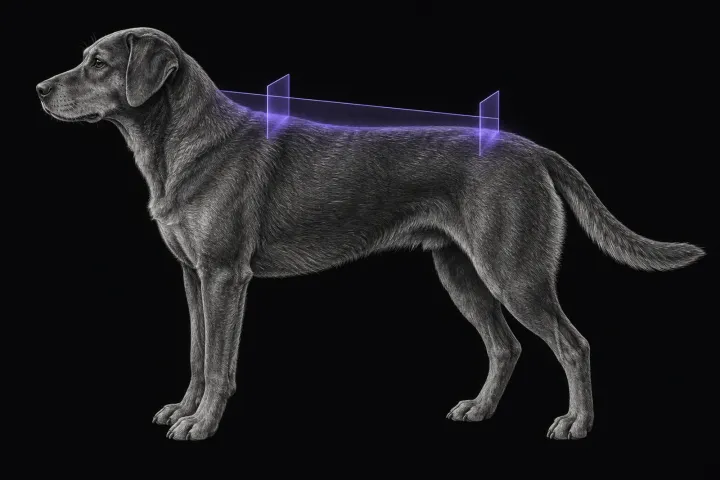
How MRI separates fibrocartilaginous embolism from acute non-compressive disc extrusion in dogs, when to scan, and which lesion measurements predict outcome.
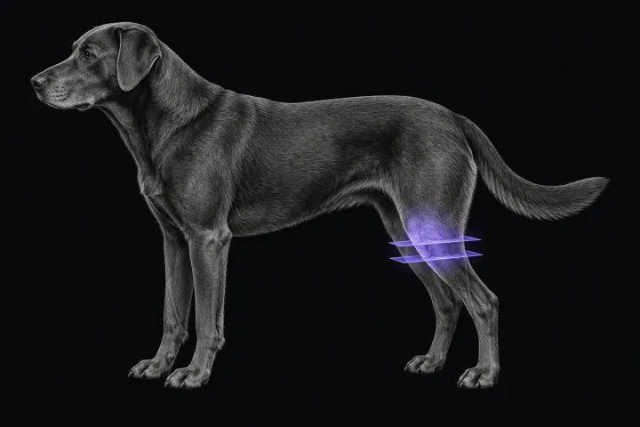
How accurately MRI shows cruciate rupture and meniscal tears in dogs, how field strength and implants affect it, and where arthroscopy still matters.
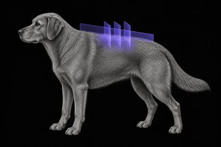
What MRI shows in dogs with acute thoracolumbar disc extrusion, how it compares with CT, and which findings matter for surgery, prognosis and myelomalacia.
How MRI separates idiopathic trigeminal neuropathy, neuritis and nerve sheath tumours in dogs with a dropped jaw or masticatory atrophy, and its pitfalls.
How radiographs, CT and MRI compare for canine discospondylitis and vertebral osteomyelitis, what MRI shows, and which mimics and follow-up limits to expect.
What brain MRI shows in cats with seizures, meningioma, lymphoma or FIP, how often the scan is normal, and what MRI cannot settle on its own.
How MRI and CT compare in cats with vestibular signs, and what MRI shows in otitis media/interna, idiopathic vestibular syndrome and central disease.
When diabetic cats need pituitary imaging for hypersomatotropism, what MRI and CT show, and why a normal-sized gland does not rule it out.
How MRI separates lymphoma, FIP myelitis, ischaemic myelopathy and disc disease in cats with myelopathy, and where CSF and biopsy are still needed.
How MRI classifies disc extrusion, protrusion and non-compressive extrusion in cats, why degeneration is often incidental, and what outcome data show.