
Case Quiz January 2026
- Patient: Canine, Kelpie Cross, 7 years, Female.
- Presenting Signs: Chronic lameness involving the pelvic limbs and hips, with a recent clinical presentation of acute spinal pain.
Case of the month: January 2026
Patient: Canine, Kelpie Cross, 7 years, Female.
History: Chronic lameness involving the pelvic limbs and hips, with a recent clinical presentation of acute spinal pain.
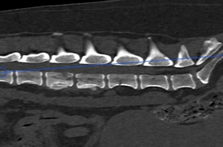
Findings: A CT study of the full spine was performed. At the L3-L4 intervertebral disc (IVD) space, there is significant bone lysis of the vertebral endplates. An ill-defined periosteal reaction is visible, predominantly on the left side of both vertebral bodies. Post-contrast images demonstrate marked tissue enhancement at the ventral aspect of the vertebral canal, localized between the L3-L4 disc space. No associated soft tissue mass is identified within the perivertebral musculature.
In the thoracic spine, there is extensive ventral new bone formation at T5-T6, accompanied by sclerosis and poor definition of the cranial endplate of T6.
Pelvis/Hips: Marked periarticular new bone formation is present at the right coxofemoral joint, associated with a shallow acetabulum and decreased femoral head coverage. Similar, though milder, changes are noted on the left.
Additional finding: A 9.5mm cholelith (gallstone) is present.
Differential Diagnoses:
- Discospondylitis (L3-L4): Highly likely due to lysis centered on the disc space and involvements of adjacent endplates.
- Spondylosis Deformans (T5-T6): Most likely cause for the thoracic bridge formation.
- Osteosarcoma: Ruled out due to the polyostotic nature (affecting two adjacent bones across a disc space) and lack of a primary bone mass.
- Infiltrative Soft Tissue Neoplasia: Ruled out due to the absence of a visible soft tissue mass on CT.
Conclusion/Diagnosis:
The primary diagnosis is Discospondylitis at L3-L4. The hallmark imaging feature confirming this is the vertebral endplate lysis centered specifically around the disc space.
Secondary findings include:
- Severe bilateral coxofemoral Degenerative Joint Disease (DJD) secondary to hip dysplasia/laxity.
- Spondylosis deformans at T5-T6.
- Cholelithiasis.
Outcome:
The recommended diagnostic plan includes blood cultures and urine culture to identify the causative infectious agent (e.g., Brucella canis, Staphylococcus spp.). Long-term antibiotic therapy is indicated, with a follow-up CT scan recommended post-treatment to confirm bone remodeling and resolution of the infectious process.
Discussion:
Discospondylitis is an infection of the intervertebral disc and the adjacent vertebral endplates. In this case, the CT was definitive in differentiating between neoplasia and infection. While primary bone tumors (like osteosarcoma) are usually monostotic and do not cross the disc space, discospondylitis is characteristically centered on the IVD. The patient’s concurrent hip dysplasia likely complicated the initial clinical assessment of lameness, highlighting the importance of advanced imaging when neurological or spinal pain is suspected.
For full case clickhere. Smarter Teleradiology. Faster Diagnosis. Better Results Join the Veheri Community! Start today: Book your DEMO