
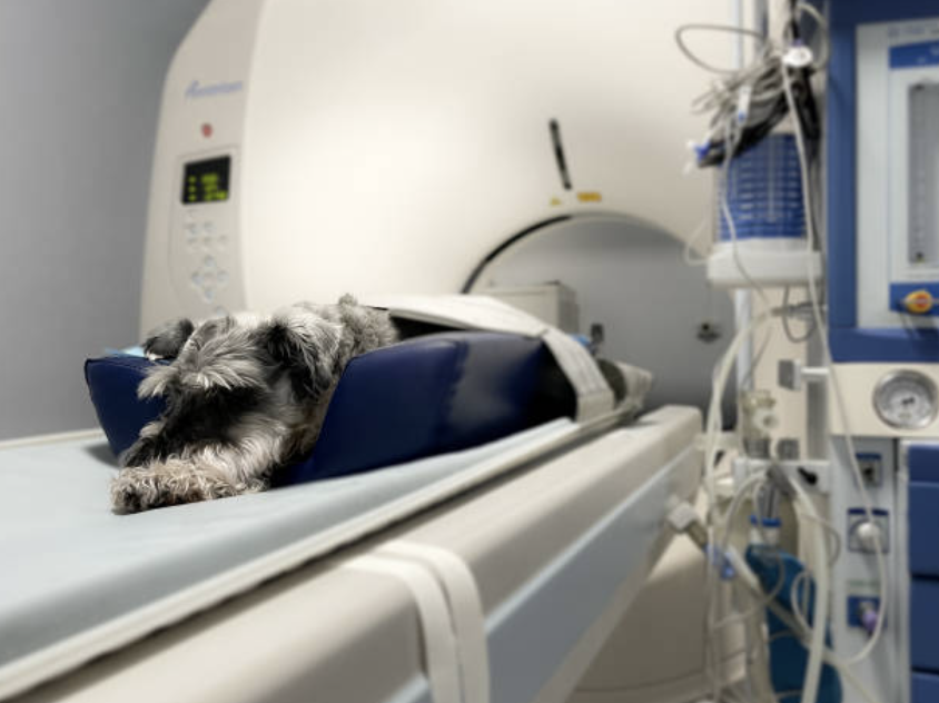
In the world of veterinary medicine, we often go looking for “The Answer” in an X-ray or a CT scan. There’s a common myth that once an image is captured, the diagnosis should be self-evident. But as any radiologist will tell you: an image is only as clear as the process behind it.
I’m thrilled to start this new series, Teleradiology Diaries with Veheri.com, where we’ll go behind the screens to talk about what actually happens when your studies hit a radiologist’s desk and how we can work together to get the best outcomes for our patients.
Precision is a Practice, Not a Guarantee
In the world of veterinary medicine, we often go looking for “The Answer” in an X-ray or a CT scan. There’s a common myth that once an image is captured, the diagnosis should be self-evident. But as any radiologist will tell you: an image is only as clear as the process behind it.
I’m thrilled to start this new series, Teleradiology Diaries with Veheri.com, where we’ll go behind the screens to talk about what actually happens when your studies hit a radiologist’s desk and how we can work together to get the best outcomes for our patients.
Moving Beyond the “Error” Narrative
We often talk about “human error” in radiology as a negative, but I prefer to look at it through the lens of Optimization. Even the most experienced board-certified radiologists are human. Interpretation is a cognitive process influenced by many factors:
- The Positioning Puzzle: Slight rotation, an uncooperative patient, or motion artefact can easily obscure subtle lesions or even mimic pathology that isn’t truly present.
- The Signal-to-Noise Challenge: Technical settings are not just buttons on a machine; they determine image quality and diagnostic value. Poor settings can leave you with a chaotic, noisy image (essentially “static”), whereas optimal settings transform that same data into a clear diagnostic tool capable of revealing crucial findings such as foreign bodies, fractures, or soft tissue abnormalities.
- The Missing Piece:A radiologist looking at a screen without clinical history is like a detective arriving at a crime scene with a blindfold on.
- Neurobias: Humans did not evolve through millions of years of selective pressure to practice radiology
The Veheri Standard: Systems Over Luck
At Veheri, we believe excellence isn’t the absence of mistakes, it’s the presence of high-standard systems. To bridge the “gap” and ensure the highest diagnostic accuracy, we focus on three pillars of Good Practice:
- Context is King: Providing a complete clinical history reduces diagnostic uncertainty by up to 50%. When we know the symptoms, we know exactly where to “zoom in.”
- Collaborative Review: Two heads are better than one. Teleradiology allows for second opinions and structured reporting that catches what a single pair of tired eyes might miss.
- Technical Synergy: High-quality interpretation starts at the table. Correct positioning and technique are the first steps in a successful diagnostic journey.
The Goal?Transparency. By acknowledging the complexities of imaging, we don’t undermine trust-we strengthen it. We aren’t just looking for “errors”; we are looking for the truth of the patient’s health.
Start today:Book your DEMOSmarter Teleradiology. Faster Diagnosis. Better Results.