
Computed Tomography (CT) is the gold standard for diagnosing complex orthopedic conditions in veterinary medicine, particularly when subtle bone changes or joint incongruities are suspected. When dealing with forelimb lameness, achieving pristine diagnostic images is heavily dependent on flawless patient positioning and precise technical execution.
This guide outlines the essential routine protocol for Elbow CT imaging to ensure maximum diagnostic resolution and accurate assessment of joint pathologies.
1. Primary Indications
An elbow CT study is most frequently indicated for diagnosing and planning treatment for:
- Elbow Dysplasia: Including fragmented medial coronoid process (FCP), ununited anconeal process (UAP), and osteochondrosis dissecans (OCD).
- Humeral Condylar Fractures / Intracondylar Fissures (HIF):Critical for surgical planning and evaluating bone stock.
2. Patient Preparation & Perfect Positioning
Proper alignment is non-negotiable for elbow CT. Because both joints are typically scanned simultaneously for comparison, asymmetry can severely compromise your diagnostic capability.
- Recumbency: Position the patient in dorsal recumbency.
- Forelimb Extension: Pull both front limbs fully in a cranial direction, ensuring the humeri are perfectly parallel to one another.
- The 110° Rule: Maintain the elbows at a strict 110° angle.
- Olecranon Alignment: Crucial Note! The olecranon must remain perfectly perpendicular to the table to avoid rotational artifact.
- Laser Alignment: Set your lateral light guide laser beam exactly at the level of the elbows.
Pro Tip: Ensure the positioning is straight and symmetrical, with minimal empty space between the patient and the lateral sides of your scan block to optimize the display field of view (DFOV).
3. Scan Parameters & Coverage
Capturing the entire joint space with high resolution requires precise anatomical landmarks for your Topogram (Scout) and Scout lines.
- Topogram: Acquire a Ventrodorsal (VD) topogram using a Top tube position to check for any patient rotation before initiating the scan.
- Scan Range: For both pre- and post-contrast series, plan your scan block to run from the mid-humerus to approximately 3 cm distal to the radial head.
Suggested Scanning Parameters
While every CT system is unique and may require alterations from your applications specialist, these baseline parameters serve as an optimal starting point:
| Parameter | Suggested Setting |
|---|---|
| kV | 100–140 kV |
| mAs | 100 mAs |
| Rotation Time | 1 sec |
| Pitch | 1.5 |
| Slice Thickness | Approx. 1.5 mm |
| Overlap / Increment | 0.25 mm (50%) |
4. Contrast Medium Protocol
While a pre-contrast scan is standard, contrast medium administration provides vital clues under specific clinical scenarios.
- IV Access: Always maintain secure I/V access throughout the procedure.
- When to use Contrast: Post-contrast scans are necessary if neoplasia, infection, or active inflammation is suspected.
- Dose & Medium: Administer $2\text{ ml/kg}$ of Iohexol ($300/350\text{ mg I/ml}$) intravenously by hand.
- Timing: Start the post-contrast scan exactly 1 minute post-injection.
5. Reconstructions to Submit
To ensure a seamless teleradiology review or internal assessment, pay close attention to your reconstruction algorithms. Note: For this protocol, submit Transverse views only!
- Pre-Contrast: Reconstruct using a Soft Tissue/Mediastinum (Soft/Medium soft) algorithm.
- Post-Contrast (If indicated): Submit two separate reconstructions:
- Osteo or Sinus (Sharp) algorithm to evaluate fine bone detail.
- Soft Tissue/Mediastinum (Soft/Medium soft) algorithm to check for contrast enhancement in surrounding soft tissues.
[Download the Complete Protocol Now]
Start today: Book your DEMO Smarter Teleradiology. Faster Diagnosis. Better Results.
Imaging guides on this topic
Browse all imaging guides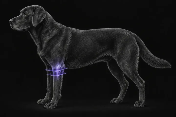
CT of the canine elbow for medial coronoid disease
When CT is worth it for medial coronoid disease in dogs, what it finds that radiographs miss, and where arthroscopy still gives the better answer.