
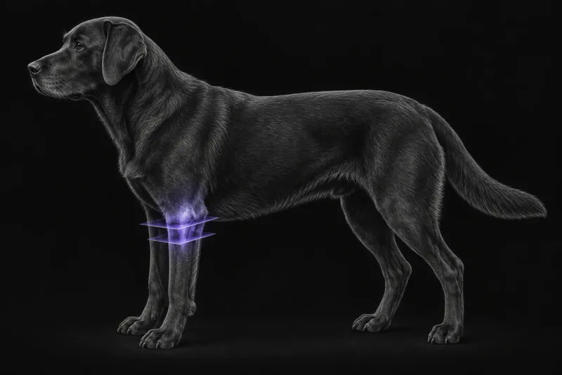
Why medial coronoid disease deserves a clear answer
Medial coronoid process disease is a common cause of thoracic limb lameness in dogs. In a retrospective study of lame large-breed dogs (Mostafa et al., Veterinary Radiology & Ultrasound, 2018), its prevalence was 20.8%, and Labrador and Golden Retrievers were the most affected purebred dogs. The authors note that CT and arthroscopy are superior to radiography for this diagnosis, while radiography remains the most available modality in practice.
Where radiographs fall short
Radiographs miss a proportion of diseased elbows, and the proportion depends on the population:
- Screening population. In 424 elbows with CT as the reference (Vali et al., Veterinary Sciences, 2025), radiography identified 17 of 26 affected joints (65%) and had a specificity of 93%. At that low prevalence (6%), the positive predictive value was 37% and the negative predictive value 98%. The authors conclude that CT is the preferred screening technique when it is available.
- Difficult cases. In dogs presented for an official second opinion (Wennemuth et al., Veterinary and Comparative Orthopaedics and Traumatology, 2020), radiography had a sensitivity of 83.6% and a specificity of 83.5% compared with CT. A false-negative radiograph was significantly associated with a fissure line on CT, and the authors strongly recommend CT in appeal cases.
- Age. In Labrador Retrievers with medial coronoid disease (Lau et al., Veterinary Surgery, 2015), radiographic sensitivity was 93.8% in dogs aged 12 months or less and 73.3% in older dogs. CT sensitivity was 93.8% and 66.7% in older dogs.
That last result is a reminder that CT is not a perfect test either. In older dogs, the most common arthroscopic finding was osteochondromalacia, a cartilage change rather than a bony fragment.
CT and arthroscopy answer different questions
- CT can disagree with arthroscopy. In 101 elbows examined with both (Moores et al., Veterinary Surgery, 2008), some CT signs were associated with arthroscopic lesions and others were not. The absence of CT signs, or of arthroscopic abnormalities, did not rule out elbow lesions.
- Cartilage needs a direct look. In elbows with medial compartment erosion (Coppieters et al., Veterinary Radiology & Ultrasound, 2016), CT and arthroscopy agreed on coronoid fragmentation but not on the subchondral defect of the humeral condyle. Some fragments seen on CT could not be found at arthroscopy. The authors conclude that estimating the extent of cartilage lesions still requires arthroscopy.
- Incongruence is method-dependent. In 24 young large-breed dogs (Griffon et al., Veterinary Surgery, 2018), radioulnar incongruence was diagnosed in 26% of elbows on radiographs, 35% on CT, 78% by CT measurement (step of 2 mm or more) and 57% at arthroscopy. Agreement with arthroscopy was poor to fair, and CT did not reliably predict the arthroscopic assessment.
In practice this usually means CT maps the bone and guides the decision to scope, while arthroscopy grades the cartilage and treats what it finds.
Elbow CT protocol at a glance
For medial coronoid disease, the acquisition choice that matters most is thin slices. In 168 CT studies of dogs that all had arthroscopy (Zweifel et al., Veterinary and Comparative Orthopaedics and Traumatology, 2020), 1-mm slices gave sharper bone contours and the highest fragment detection rate, and slices of 2 mm or more significantly affected fragment detection.
What the radiologist looks at
The radiologist weighs each CT sign against what the evidence says it predicts at arthroscopy:
| Finding on CT | What the evidence shows |
|---|---|
| Fragment of the medial coronoid process | Associated with a displaced fragment and with cartilage erosion of the coronoid and humeral condyle at arthroscopy (Moores et al.) |
| Fissure line | Significantly associated with a false-negative radiograph (Wennemuth et al.) |
| Irregular radial incisure of the ulna | Associated with cartilage erosion of the medial coronoid process (Moores et al.) |
| Osteophytes | Associated with an abnormal arthroscopy; osteophyte grade correlated moderately with medial compartment cartilage erosion grade (rs = 0.44–0.48) (Moores et al.) |
| Advanced medial compartment erosion | Periarticular osteophytosis in 100%, abnormal coronoid shape and a subchondral bone defect of the medial humeral condyle in 96.4% of 56 elbows (Coppieters et al.) |
Reading is also partly subjective. In a study of eight observers (Shimizu et al., Journal of Small Animal Practice, 2015), agreement was fair to moderate for calling medial coronoid disease, moderate to almost perfect for fragmentation, and poor for grading sclerosis.
What your report will include
Every Veheri case is read by a radiologist or neurologist, and when you order you can choose Diplomate-only reporting or the full specialist panel. You can chat directly with the specialist assigned to your case, and a simplified pet owner report helps you explain the findings to the owner.
When you take the report forward:
- A CT without signs does not close the question: it did not rule out elbow lesions.
- Cartilage damage is best graded at arthroscopy, because the extent of cartilage lesions still requires arthroscopy.
- Incongruence on CT should be read with care, because CT did not reliably predict the arthroscopic assessment.
- Older dogs with a negative CT may still have cartilage disease, since CT sensitivity was lower in dogs over 12 months.
- Planning: it is worth discussing CT and arthroscopy with the surgeon before the first anaesthetic.
What to include when you request an elbow CT report
- signalment: age, breed and sex
- weight, and remember that sensitivity of both radiographs and CT differs with age
- duration of lameness, which limb, and response to rest or analgesia
- whether the scan is for a lame dog, breeding screening or an appeal
- previous radiographs and their reports, so the radiologist can compare
- any previous elbow surgery or arthroscopy
How Veheri can help
Veheri’s radiologists report veterinary CT studies remotely for clinics, and we offer expert support for CT protocols, positioning and acquisition. For the practical side of scanning, see mastering veterinary elbow CT and why contrast-enhanced CT helps limping dogs.
Sources
Each claim in this guide links to its source. The full citations:
- Mostafa A, Nolte I, Wefstaedt P. The prevalence of medial coronoid process disease is high in lame large breed dogs and quantitative radiographic assessments contribute to the diagnosis. Vet Radiol Ultrasound. 2018;59(5):516-528. doi:10.1111/vru.12632
- Vali Y, Villamonte Chevalier A, van Bree H, et al. Cross-modality diagnostic agreement in canine medial coronoid disease in a screening population: radiographs vs. CT in 424 elbow joints. Vet Sci. 2025;12(9):883. doi:10.3390/vetsci12090883
- Wennemuth J, Tellhelm B, Eley N, et al. Computed tomography enhances diagnostic accuracy in challenging medial coronoid disease cases: an imaging study in dog breeding appeal cases. Vet Comp Orthop Traumatol. 2020;33(5):356-362. doi:10.1055/s-0040-1714299
- Lau SF, Theyse LF, Voorhout G, et al. Radiographic, computed tomographic, and arthroscopic findings in Labrador Retrievers with medial coronoid disease. Vet Surg. 2015;44(4):511-20. doi:10.1111/j.1532-950X.2014.12291.x
- Moores AP, Benigni L, Lamb CR. Computed tomography versus arthroscopy for detection of canine elbow dysplasia lesions. Vet Surg. 2008;37(4):390-8. doi:10.1111/j.1532-950X.2008.00393.x
- Coppieters E, Van Ryssen B, van Bree H, et al. Computed tomographic findings in canine elbows arthroscopically diagnosed with erosion of the medial compartment: an analytical method comparison study. Vet Radiol Ultrasound. 2016;57(6):572-581. doi:10.1111/vru.12425
- Zweifel RT, DiDonato P, Hartmann A, et al. Improved computed tomography accuracy with a 1-mm versus 2- or 3-mm slice thickness for the detection of medial coronoid disease in dogs. Vet Comp Orthop Traumatol. 2020;33(1):45-50. doi:10.1055/s-0039-3399524
- Shimizu N, Warren-Smith CM, Langley-Hobbs SJ, et al. Inter- and intraobserver agreement in interpretation of CT features of medial coronoid process disease. J Small Anim Pract. 2015;56(12):707-13. doi:10.1111/jsap.12411
- Griffon DJ, Mostafa AA, Blond L, et al. Radiographic, computed tomographic, and arthroscopic diagnosis of radioulnar incongruence in dogs with medial coronoid disease. Vet Surg. 2018;47(3):333-342. doi:10.1111/vsu.12783
Related reading
More imaging guides
All imaging guides
CT of adrenal masses in dogs
What contrast CT shows in a dog with an adrenal mass, from caval invasion and adhesions to tumour type, and where its accuracy runs out before adrenalectomy.

Brain CT in dogs when MRI is not available
What brain CT can and cannot detect in dogs when MRI is not available, for masses, stroke, inflammation and head trauma, and when MRI or CSF is still needed.
CT of the canine gastrointestinal tract in the acute abdomen
When abdominal CT adds to radiographs and ultrasound in vomiting dogs with suspected obstruction, foreign body or perforation, and what CT can and cannot show.