
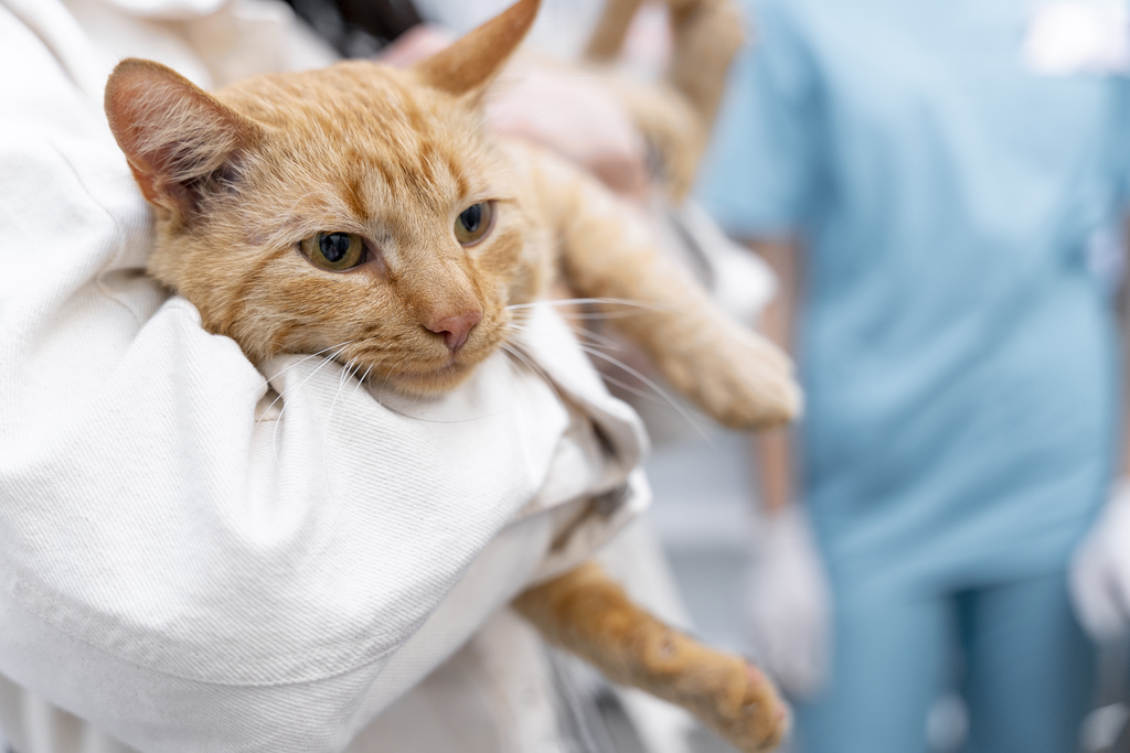
In an emergency, speed and precision aren’t just goals—they are lifesavers. While traditional imaging like ultrasound and X-rays are common first steps, the Emergency CT provides a level of detail that is unmatched for critical cases.
The Golden Hour: The Crucial Role of Emergency CT in Veterinary Practice
In an emergency, every second counts. Rapid, targeted diagnostics are the backbone of effective therapy planning and prognostic decision-making. While ultrasound (AFAST/TFAST) and radiographs are traditional first steps, the Emergency CT has emerged as a game-changer for critical patients, offering a level of detail that traditional imaging simply cannot match.
Why Choose CT in an Emergency?
Historically, CT was reserved for elective procedures due to anesthesia concerns and limited availability. Today, with faster scanning times and the rise of teleradiology on platforms like Veheri, emergency CT is more accessible than ever. It is particularly superior for:
- Precision: Avoiding the superimposition of structures seen in X-rays.
- Comprehensive Overviews: Surveying multiple body systems in minutes (essential for polytrauma).
- Surgical Planning: Identifying the exact location of hemorrhages, fractures, or foreign bodies before the first incision.
READ ALSO: Veterinary CT: WhatIt Is, When Pets Need It, Safety, and Costs
Patient Stabilization: The Priority Before the Scan
No matter how advanced the technology, clinical stabilization always takes priority over imaging. Before moving a patient to the CT suite, the ASA status must be addressed:
- Respiratory Support: Oxygenation and stabilization of pleural diseases (e.g., thoracentesis for dyspnea).
- Cardiovascular Support: Addressing shock via infusions or transfusions.
- Analgesia: Ensuring the patient is comfortable enough for positioning.
Clinical Note: If using contrast media, ensure the patient is well-hydrated to mitigate the risk of acute renal insufficiency, especially in hemodynamically unstable patients.
READ ALSO:When is a CT scan indicated in veterinary medicine?
“Awake” vs. “Anaesthetized” CT
While general anesthesia is preferred for optimal positioning and motion-free images (especially for thoracic apnea scans), it isn’t always an option.
- Anaesthesia CT: The gold standard for image quality. Allows for controlled ventilation and precise slicing.
- Awake CT: Indicated for highly unstable patients, comatose patients, or those with severe polytrauma where anesthesia is contraindicated. Using positioning aids like “cat baskets” or blankets can help stabilize the patient during the rapid scan.
Top Indications for Emergency CT
1. Traumatic Brain Injury (TBI)
CT is the method of choice for assessing intracranial pressure and hemorrhage.
- The Koret CT Score (KCTS): A valuable prognostic index for dogs and cats after head trauma. While KCTS helps predict survival, it should always be used alongside clinical progression rather than as the sole factor for euthanasia decisions.
2. Thoracic & Vascular Emergencies
- Pulmonary Thromboembolism & Lobe Torsion: CT is often the only reliable method for a definitive diagnosis.
- Respiratory Distress: While awake CT is difficult due to motion artifacts, a rapid sedated scan can clarify complex underlying pathology.
3. Polytrauma and Penetrating Injuries
Whether it’s a “Big Dog-Little Dog” attack or a high-velocity impact, CT allows for the rapid assessment of:
- Impalements & Bite Injuries: Determining the depth of involvement and surgical margins.
- Spinal Trauma: Detecting vertebral fractures and luxations that are easily missed on plain films.
Conclusion: Targeted Diagnostics
The goal of an emergency CT is not to “see what we find,” but to answer specific clinical questions. Focus on the region of interest to keep anesthesia time to a minimum. By integrating CT into your emergency workflow and utilizing Veherifor express reporting and specialist collaboration, you can significantly improve patient outcomes and provide clearer prognostic data to owners.
Join the global imaging community: Have a challenging emergency case? Get your account and upload your first case at Diagnostics.veheri.com to discuss findings with experts worldwide and explore the future of veterinary diagnostics.
Start today: Book your DEMO Smarter Teleradiology. Faster Diagnosis. Better Results.
Imaging guides on this topic
Browse all imaging guides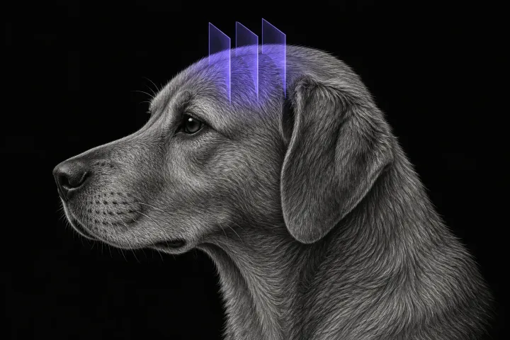
Brain CT in dogs when MRI is not available
What brain CT can and cannot detect in dogs when MRI is not available, for masses, stroke, inflammation and head trauma, and when MRI or CSF is still needed.
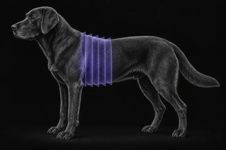
CT pulmonary angiography for suspected pulmonary thromboembolism in dogs
When to request CT pulmonary angiography for suspected pulmonary thromboembolism in dogs, what a positive scan looks like, and why technique decides the answer.
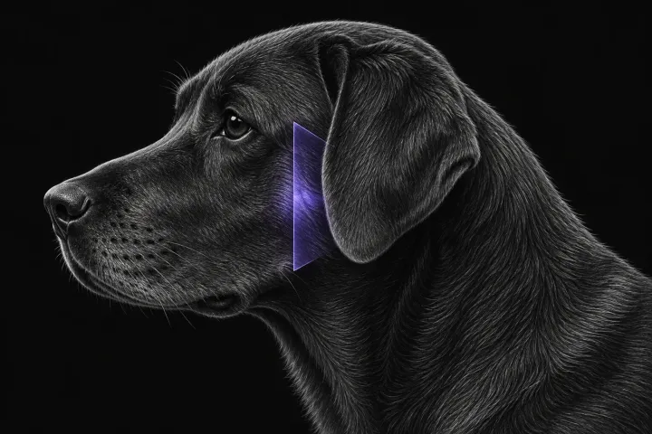
CT of the canine temporomandibular joint
When CT of the canine temporomandibular joint is indicated, what it shows in fracture, ankylosis, dysplasia and osteoarthritis, and where it falls short.