
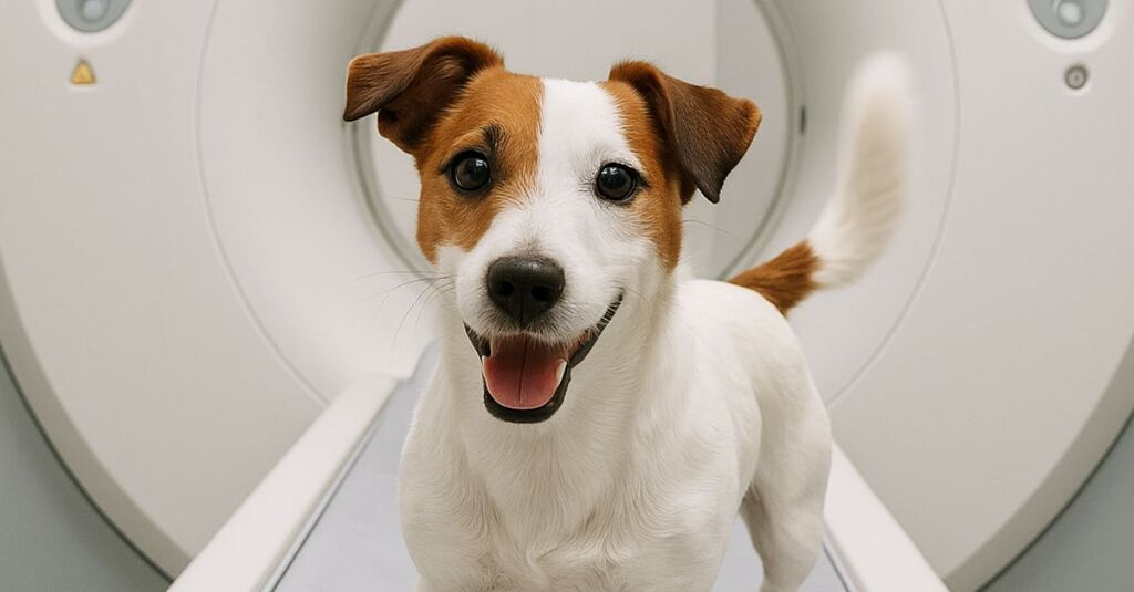
Veterinary CT provides rapid, high‑detail imaging that guides diagnosis and treatment planning across species. With appropriate anesthesia and safety protocols, CT is a mainstay of modern care, from trauma and oncology to dentistry - worldwide.
If you’re wondering what a veterinary CT is and whether your pet needs one: a CT scan uses X-rays to create detailed cross-sectional images that help vets diagnose issues in the head, chest, abdomen, spine, and limbs, often under brief anesthesia.
Below, you’ll find what veterinary CT is, when it’s recommended, how it compares with X-ray and MRI, anesthesia and safety, typical costs (globally variable), and what to expect.
What is veterinary CT?
Computed tomography (CT) uses an X‑ray tube and detectors that rotate around the patient to produce thin “slices.” These can be reconstructed into high‑detail 3D views of bones, soft tissues, and blood vessels. The resolution and speed make CT a cornerstone of modern veterinary diagnostics across species and body systems.
“Computed Tomography (CT) imaging uses X-rays in conjunction with computing algorithms to image the body.” American College of Veterinary Radiology via ACVR
When is a veterinary CT recommended?
CT is commonly used for nasal disease, head and jaw disorders, ear bulla disease, dental/oral conditions, thoracic masses, lung metastasis, portosystemic shunts, abdominal masses, complex fractures, elbow dysplasia, spinal disease, and pre‑operative or radiation planning. Referral hospitals often use contrast-enhanced CT angiography for vascular mapping and oncology staging.
A CT Scan is always recommended based on the professional judgment of a qualified veterinarian. If your veterinarian suggests a CT, you can trust in the expertise and commitment to providing the best possible care for your pet.
For clinics: CT by body region
If you run CT in your practice or refer cases for it, these guides go deeper on specific body regions, with protocols and cited evidence:
- CT of the canine nasal cavity for chronic nasal disease
- CT of the feline middle ear
- Mastering veterinary elbow CT: a comprehensive protocol guide
- Mastering veterinary spinal CT: a comprehensive protocol guide
- Optimizing veterinary CT: abdominal routine protocols
For brain and spinal cord questions, MRI is usually the better tool: see brain MRI for dogs with seizures. Veheri’s radiologists report veterinary CT and MRI studies remotely for clinics.
Veterinary CT vs. X‑ray vs. MRI
Modality choice depends on the clinical question:
-
CT vs X-ray:CT delivers far more detail, cross‑sections, and 3D reconstructions. It’s excellent for complex bone, nasal/sinus disease, thoracic evaluation (including metastasis), and surgical/radiation planning.
-
CT vs MRI: MRI is superior for brain and spinal cord and many soft‑tissue or neurologic conditions. CT is faster and typically better for bone, lungs, mineralization, and intricate bony anatomy.
For dentistry and oral surgery, cone beam CT (CBCT) is increasingly used to visualize teeth and jaws with high spatial resolution.
Anesthesia, safety, and what to expect
Most small animals must stay completely still for a CT, which is why sedation or general anesthesia is commonly used. Modern scanners are fast, reducing anesthesia time. Your team will screen for risks, monitor throughout, and follow radiation safety protocols; benefits generally outweigh risks when CT is appropriately indicated.
“Dogs will be given general anesthesia for CTs or MRIs to make sure they do not move during the procedure.” Charlotte Hacker, PhDviaPetMD
How much does a veterinary CT cost?
Costs vary widely worldwide by region, facility, contrast use, and add‑on diagnostics.
“Selecting the best provider for a veterinary CT system is a critical decision that impacts your clinic’s diagnostic capabilities, workflow, and long-term costs. To make*an informed choice,it’s essential to consider not just the price, but the full spectrum of services and support that come with your investment.” -*How to Choose the Right Veterinary CT Modailty: 8 Key Considerations for Your Practice
Consider pre-anesthetic bloodwork, anesthesia time, radiologist interpretation, and potential same‑day follow‑up imaging. Wherever you are, ask your local referral hospital for a written estimate detailing these components.
Innovations you’ll see worldwide
CT continues to advance. Photon‑counting CT—long established in human medicine—is entering veterinary centers and may improve resolution and dose efficiency.
“Photon-counting CT is the newest CT technology, with fewer than 50 scanners in clinical use, and this is the first installed in veterinary medicine,” said Peter Scrivani ’89, D.V.M. ’93. - Peter V. Scrivani, DVM, ACVRviaCornell University College of Veterinary Medicine
For oncology, advanced centers integrate CT data into radiation planning—including stereotactic radiosurgery.
Preparing your pet and timeline
A typical CT visit can take a few hours from check‑in to discharge.
Expect:
- Pre‑anesthetic exam and bloodwork (as indicated), IV catheter placement, and possibly contrast dye
- Short anesthesia or deep sedation, CT acquisition, and recovery monitoring
- A radiologist’s interpretation and communication back to your primary vet/team
Owner tips:
- Follow fasting instructions to reduce anesthesia risk.
- Bring prior imaging and records.
- Ask if contrast will be used and about hydration afterward.
Smarter Teleradiology. Faster Diagnosis. Better Results
Join the Veheri Community!
Start today: Book your DEMO