
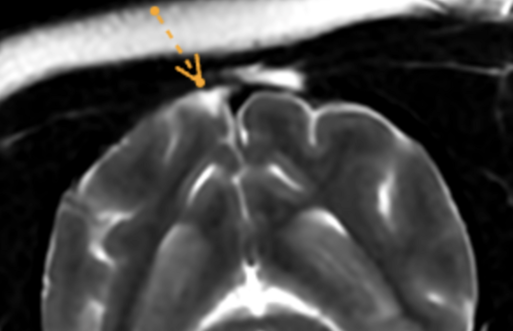
In the past, many veterinary neurological conditions were categorized as “idiopathic”—an elaborate way of saying we didn’t know the cause. Today, the landscape has shifted. With the integration of Advanced Veterinary Neurology Reporting and high-field MRI and CT, we are now able to visualize the invisible.
In the past, many veterinary neurological conditions were categorized as “idiopathic”—an elaborate way of saying we didn’t know the cause. Today, the landscape has shifted. With the integration of Advanced Veterinary Neurology Reporting and high-field MRI and CT, we are now able to visualize the invisible.
When a pet presents with neurological signs, imaging is the bridge between a mystery and a management plan. Here are the most common neurological diseases currently diagnosed through specialist-led diagnostic imaging.
1. Intervertebral Disc Disease (IVDD)
IVDD remains the most frequent reason for spinal imaging in dogs. While X-rays can suggest a narrow disc space, they cannot see the disc material itself or the bruising of the spinal cord.
- The Imaging Edge: MRI is the gold standard for identifying the type of herniation (Hansen Type I vs. Type II) and the exact degree of spinal cord compression.
- Clinical Impact: This allows for precise surgical planning or the decision to manage the case medically with confidence.
2. Neoplasia (Brain and Spinal Tumors)
Tumors of the nervous system—such as Meningiomas, Gliomas, or Nerve Sheath Tumors—are impossible to diagnose via bloodwork or physical exams.
- The Imaging Edge: Contrast-enhanced MRI allows ECVN Specialists to see the margins of a tumor, its vascularity, and its effect on surrounding brain tissue.
- Clinical Impact: Early detection via imaging can make the difference between a terminal prognosis and successful surgical removal or radiation therapy.
3. Syringomyelia and Chiari-like Malformation (CM)
Common in toy breeds like Cavalier King Charles Spaniels, this condition involves a structural mismatch between the skull and the brain, leading to fluid-filled cavities (syrinxes) in the spinal cord.
- The Imaging Edge: Only MRI can visualize the flow of cerebrospinal fluid and the presence of a syrinx.
- Clinical Impact: Imaging prevents these pets from being misdiagnosed with “allergies” or “skin issues” due to their phantom scratching behavior.
4. Inflammatory CNS Diseases (GME/MUE)
Meningoencephalitis of Unknown Etiology (MUE) is a group of non-infectious inflammatory diseases where the immune system attacks the brain.
- The Imaging Edge: MRI reveals characteristic “lesions” within the brain parenchyma that are invisible to CT scans and X-rays.
- Clinical Impact: Rapid diagnosis allows for the immediate start of immunosuppressive therapy, which is vital for survival.
5. Vascular Accidents (Strokes)
Just like humans, animals suffer from ischemic (clot) or hemorrhagic (bleed) strokes.
- The Imaging Edge: Advanced MRI sequences (like DWI and FLAIR) can identify a stroke within hours of the event.
- Clinical Impact: Identifying a stroke prevents unnecessary surgery and allows the focus to shift toward supportive care and identifying underlying causes like hypertension.
The Power of Specialized Interpretation: Beyond the Image
Acquiring a high-quality scan is only the first step; unlocking the clinical narrative within those images is where the true diagnosis lives. CT & MRI Interpretation by ECVN Specialists ensures that subtle, life-altering pathologies—such as lacunar infarcts, early-stage neuroinflammation, or nerve root entrapment—are identified with precision.
Why Expert Reporting is a Clinical Game-Changer:
- Differential Precision: Expertly differentiating between neoplastic, inflammatory, and infectious processes (e.g., tumor vs. abscess) to avoid contraindicated treatments.
- From “Finding” to “Action”: We don’t just describe lesions; we provide a therapy-focused roadmap, translating complex pixels into actionable medical or surgical steps.
- Data-Driven Prognosis: Empowering you to offer pet owners clear, evidence-based expectations regarding recovery and long-term quality of life.
Elevate Your Diagnostic Confidence
In neurology, the margin for error is slim. Don’t leave your most complex cases to chance. Our ECVN-certified neurologists deliver comprehensive, clinically correlated reports designed to integrate seamlessly into your workflow.
Partner with us to transform your diagnostic process. Contact us today to submit your first case or to start a trial of our reporting service. Experience the clinical impact of board-certified expertise on your patient outcomes.
Start today:Book your DEMOSmarter Teleradiology. Faster Diagnosis. Better Results.
Imaging guides on this topic
Browse all imaging guides
Brain CT in dogs when MRI is not available
What brain CT can and cannot detect in dogs when MRI is not available, for masses, stroke, inflammation and head trauma, and when MRI or CSF is still needed.
MRI of the inner ear and vestibular system in dogs
How reliably the neurological exam separates central from peripheral vestibular disease in dogs, and what MRI shows in otitis interna and idiopathic cases.
MRI of the trigeminal nerve in dogs with a dropped jaw or masticatory atrophy
How MRI separates idiopathic trigeminal neuropathy, neuritis and nerve sheath tumours in dogs with a dropped jaw or masticatory atrophy, and its pitfalls.