
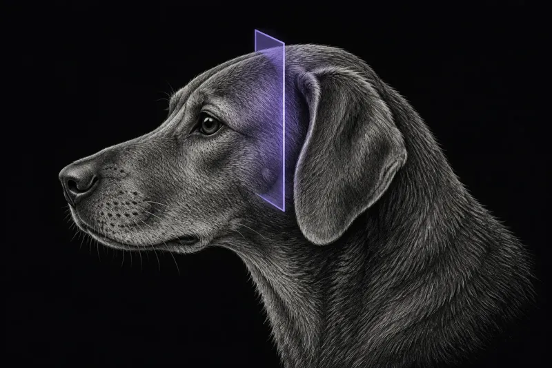
Two presentations, different likely causes
Trigeminal nerve disease in dogs usually presents in one of two ways.
A dropped jaw. In 29 dogs unable to close the mouth (Mayhew et al., Journal of the American Animal Hospital Association, 2002) because of flaccid paralysis or paresis of the muscles supplied by the mandibular branch, 26 were diagnosed with idiopathic trigeminal neuropathy on the basis of complete resolution. Mean time to recovery was 22 days. Lymphosarcoma, Neospora caninum infection and severe polyneuritis were found at necropsy in the other three.
Unilateral masticatory muscle atrophy. In 63 dogs referred for this sign and imaged with MRI (Milodowski et al., Journal of Veterinary Internal Medicine):
- a presumptive trigeminal nerve sheath tumour was diagnosed in 30 (47.6%)
- another extra-axial mass in 13 (20.6%)
- no causative lesion in 18 (28.6%)
What separates the causes
| Group | Share | Laterality |
|---|---|---|
| Idiopathic trigeminal neuropathy | 53% | 22 bilateral, 4 unilateral |
| Neoplasia | 31% | All unilateral |
| Neuritis | 16% | 1 bilateral, 7 unilateral |
- Bilateral signs gave a predicted probability of idiopathic trigeminal neuropathy of 95.7%.
- Unilateral signs and sensory loss were associated with neoplasia or neuritis.
- Age. Dogs with neoplasia were older than dogs with neuritis or idiopathic neuropathy.
- Central deficits can be absent even with marked brainstem involvement by a tumour.
flowchart TD
  accTitle: Bilateral or unilateral trigeminal signs?
  accDescr: From a blinded MRI review of 49 dogs. Bilateral signs made idiopathic trigeminal neuropathy very likely, while unilateral signs and sensory loss were associated with neoplasia or neuritis.
  start["Dropped jaw or masticatory atrophy"] --> side{{"Signs bilateral?"}}
  side -- "Yes" --> itn["Idiopathic neuropathy most likely"]
side -- "No" --> mass["Neoplasia or neuritis more likely"]In the unilateral atrophy series, 25 of 30 dogs with a presumptive nerve sheath tumour (83%) and all 13 dogs with another extra-axial mass had other neurological deficits. In the 18 dogs without an MRI lesion, neurological signs progressed in only one.
Trigeminal nerve MRI protocol at a glance
The study is a brain MRI that follows both trigeminal nerves from the brainstem to their branches, with pre- and post-contrast imaging. In six dogs with histologically confirmed disease (Schultz et al., Veterinary Radiology & Ultrasound, 2007), lesions were assessed on T1-weighted, T2-weighted and proton density images, and all enhanced after contrast. The field of view should include the middle ears, because effusion there can be part of the picture.
What the radiologist looks at
- Neuritis or tumour? In the six-dog study:
- neuritis showed diffuse enlargement of the nerve without a mass, isointense to brain on T1-weighted and proton density images and iso- or hyperintense on T2-weighted images
- a nerve sheath tumour formed a solitary or lobulated mass displacing adjacent neuropil, isointense to brain on T1-weighted, T2-weighted and proton density images
- all dogs had atrophy of the temporalis and masseter muscles, with increased signal on T1-weighted images
- Extent of a tumour. In 27 dogs with imaging-diagnosed trigeminal nerve sheath tumours (Swift et al., Veterinary Radiology & Ultrasound, 2017), all had disease extending centrally to the level of the brainstem. The mandibular branch was most often involved (26 dogs), then the maxillary (22) and ophthalmic (10). Presenting signs were masticatory muscle atrophy (26), intracranial neurological signs (13) and ocular disease (12).
- Normal nerves enhance. In 42 dogs with otherwise normal brain MRI and no clinical trigeminal disease (Pettigrew et al., Veterinary Radiology & Ultrasound, 2009), the whole nerve enhanced in 39 and the region of the ganglion in all 42, with intensity no greater than the pituitary gland. Enhancement alone is not proof of disease.
- Bilateral disease has no normal side to compare. In 137 dogs without trigeminal disease (Swain et al., Frontiers in Veterinary Science, 2020), nerve-to-brain height and width ratios measured on post-contrast 3D T1-weighted reconstructions gave reference intervals of 0.07–0.11 and 0.08–0.13. The ratios varied with body weight and skull type, so the authors call for further work before relying on them.
- Middle ear effusion may be neurogenic. The mandibular branch supplies the tensor veli
palatini muscle, which opens the auditory tube. Ipsilateral effusion was found in:
- 6 of 18 dogs with trigeminal dysfunction (Kent et al., Journal of Veterinary Internal Medicine, 2013)
- 63% of 19 dogs with trigeminal mass lesions (Wessmann et al., Veterinary Record, 2013), where it went with more severe masticatory muscle loss
- only dogs with neoplasia (33%) in the 49-dog series, as non-infectious effusion
- 18 of 30 dogs with a presumptive tumour in the unilateral atrophy series
What your report will include
Every Veheri case is read by a radiologist or neurologist, and when you order you can choose Diplomate-only reporting or the full specialist panel. Neurology reporting covers CT and MRI. You can chat directly with the specialist assigned to your case, and a simplified pet owner report helps you explain the findings to the owner.
When you take the report forward:
- Most diagnoses are presumptive. In the unilateral atrophy series, the 49-dog series and the 27-dog tumour series, most diagnoses were based on MRI, without histology.
- A normal MRI does not end the work-up. No cause was found in 18 dogs in the unilateral atrophy series.
- Extent towards the brainstem matters for treatment planning, because all 27 tumours in one series reached that level.
- Talking to the owner: dogs with mass lesions were more likely to deteriorate or be euthanised, while a dropped jaw from idiopathic neuropathy resolved completely, with a mean recovery time of 22 days.
What to include when you request an MRI report
- signalment: age, breed and sex
- whether the jaw can close, and whether signs are unilateral or bilateral
- onset and duration: sudden dropped jaw or gradual muscle atrophy
- facial sensation, palpebral and corneal reflexes
- other cranial nerve deficits, Horner syndrome, head tilt or ocular disease
- otoscopic findings, so a middle ear effusion can be interpreted
How Veheri can help
Veheri provides veterinary teleradiology and teleneurology and reports brain MRI studies remotely for clinics. For masticatory muscle disease, see MRI of the canine masticatory muscles, and for other presentations, when veterinary MRI is the right move.
Sources
Each claim in this guide links to its source. The full citations:
- Mayhew PD, Bush WW, Glass EN. Trigeminal neuropathy in dogs: a retrospective study of 29 cases (1991-2000). J Am Anim Hosp Assoc. 2002;38(3):262-70. doi:10.5326/0380262
- Milodowski EJ, Amengual-Batle P, Beltran E, et al. Clinical findings and outcome of dogs with unilateral masticatory muscle atrophy. J Vet Intern Med. 33(2):735-742 (published online 2018). doi:10.1111/jvim.15373
- Musso C, Le Boedec K, Gomes E, Cauzinille L. Diagnostic values of clinical and magnetic resonance findings in presumptive trigeminal neuropathy: 49 dogs. J Am Anim Hosp Assoc. 2020;56(2):106-113. doi:10.5326/JAAHA-MS-6997
- Schultz RM, Tucker RL, Gavin PR, et al. Magnetic resonance imaging of acquired trigeminal nerve disorders in six dogs. Vet Radiol Ultrasound. 2007;48(2):101-4. doi:10.1111/j.1740-8261.2007.00212.x
- Swift KE, McGrath S, Nolan MW, et al. Clinical and imaging findings, treatments, and outcomes in 27 dogs with imaging diagnosed trigeminal nerve sheath tumors: a multi-center study. Vet Radiol Ultrasound. 2017;58(6):679-689. doi:10.1111/vru.12535
- Pettigrew R, Rylander H, Schwarz T. Magnetic resonance imaging contrast enhancement of the trigeminal nerve in dogs without evidence of trigeminal neuropathy. Vet Radiol Ultrasound. 2009;50(3):276-8. doi:10.1111/j.1740-8261.2009.01533.x
- Swain CE, Cherubini GB, Mantis P. Low field MRI measurements of the normal canine trigeminal nerve. Front Vet Sci. 2020;7:274. doi:10.3389/fvets.2020.00274
- Kent M, Glass EN, de Lahunta A, et al. Prevalence of effusion in the tympanic cavity in dogs with dysfunction of the trigeminal nerve: 18 cases (2004-2013). J Vet Intern Med. 2013;27(5):1153-8. doi:10.1111/jvim.12140
- Wessmann A, Hennessey A, Goncalves R, et al. The association of middle ear effusion with trigeminal nerve mass lesions in dogs. Vet Rec. 2013;173(18):449. doi:10.1136/vr.101817
Related reading
More imaging guides
All imaging guides
MRI of the canine brachial plexus for peripheral nerve sheath tumours
How MRI finds peripheral nerve sheath tumours of the canine brachial plexus, what the radiologist looks for, and what imaging can say about grade and extent.
Brain MRI for dogs with seizures
Which dogs with seizures need brain MRI, what the evidence says about diagnostic yield, what the radiologist looks for and what your report will include.
MRI of the canine cervical spine for cervical spondylomyelopathy
How MRI characterises cervical spondylomyelopathy in dogs, what traction and flexion-extension studies add, and why compression alone does not prove the cause.