
Case Quiz November 2025
- Patient: 6-year-old British Shorthair cat
- Presenting Signs: Tachypnea and dyspnea
Signalment and History:
6-year-old British Shorthair, presented due to tachypnea and dyspnea.
Radiographic Description:
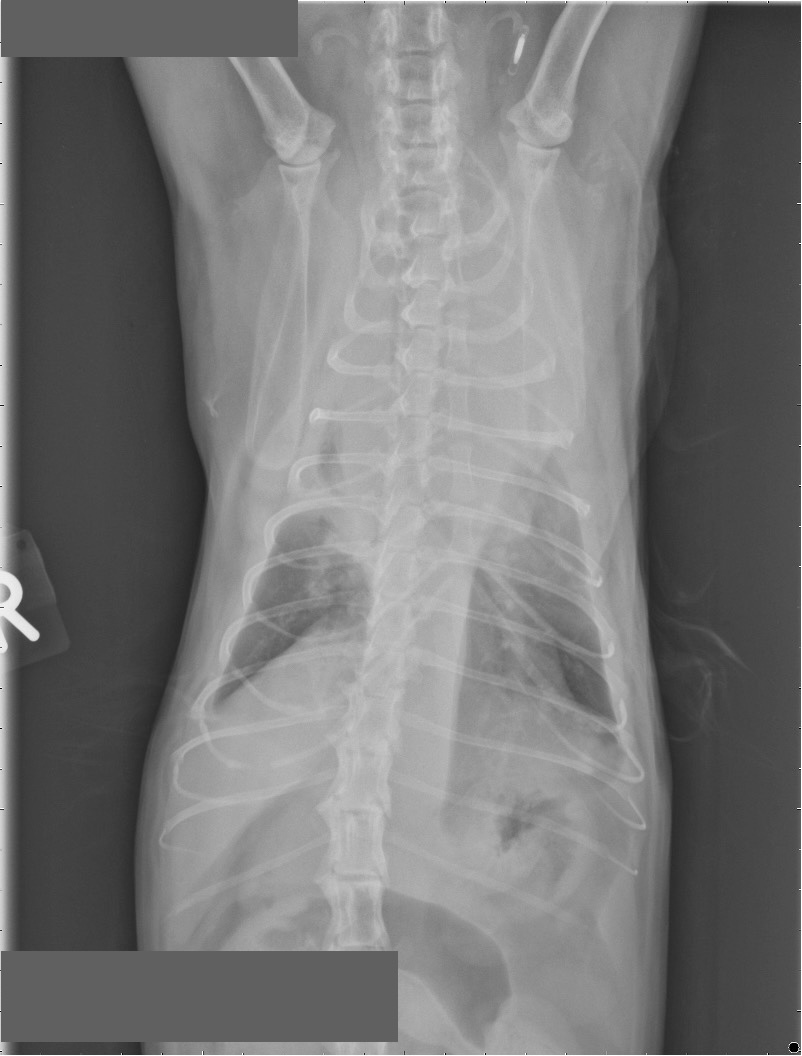
A moderate amount of soft tissue opaque pleural effusion is visible bilaterally. In the cranial mediastinum, a soft tissue opaque mass is present, causing caudal displacement of the cardiac silhouette, and caudal shift of the cranial lung margins.
The cardiac silhouette shows partial contour effacement due to the mediastinal mass and bilateral pleural
effusion; the visible portions, however, appear within normal limits.
In the ventilated caudal lung fields, a mildly increased, diffusely unstructured interstitial pattern is visible. The
visible pulmonary vessels appear unremarkable, and the trachea shows uniform diameter and course.
The visible cranial abdomen demonstrates adequate serosal contrast. The surrounding musculoskeletal structures are unremarkable.
Diagnosis:
The findings are consistent with a cranial mediastinal mass, most likely neoplastic such as mediastinal
lymphoma or thymoma. The present bilateral pleural effusion is considered secondary to the mediastinal mass. Differential diagnoses also include ectopic thyroid tissue or sternal lymphadenopathy as less likely causes.
Outcome:
Following further diagnostics (fine-needle aspirate and cytopathology of the mediastinal mass), lymphoma was confirmed. The cat received chemotherapy, under which the pleural effusion completely resolved. In the
following weeks, the cat showed marked clinical improvement and stable remission.
Discussion:
Lymphomas are the most commonly diagnosed tumors in cats, accounting for approximately 30% of all
malignant diseases. Historically, prior to widespread FeLV vaccination, around 70% of cats with lymphoma
were FeLV-positive, and mediastinal lymphomas involving the thymus as well as mediastinal and sternal lymph nodes were common. After the introduction of FeLV vaccination, both FeLV prevalence and the proportion of mediastinal lymphoma cases decreased significantly; currently, they account for <15–25% of feline lymphoma cases. Various chemotherapy protocols, such as COP (cyclophosphamide, vincristine, prednisone) and Madison–Wisconsin (doxorubicin, vincristine, cyclophosphamide, prednisone), demonstrate overall high response rates in extranodal lymphomas (87% overall response; median survival times 112–171 days), although cats with mediastinal lymphoma have been underrepresented in large studies. Current research aims to describe signalment, retroviral status, treatment response, and survival times in cats with mediastinal lymphoma and to identify potential prognostic factors.
Get access to the full case study here: Case of the Month – November 2025
Further reading:
Trisha J. Oura, Philip E. Hamel, Samuel H. Jennings, Perry J. Bain, Dennis E. Jennings, John Berg;
Radiographic Differentiation of Cranial Mediastinal Lymphomas from Thymic Epithelial Tumors in Dogs and
Cats. J Am Anim Hosp Assoc 1 July 2019; 55 (4): 187–193. doi: https://doi.org/10.5326/JAAHA-MS-6907
Fabrizio F, Calam AE, Dobson JM, Middleton SA, Murphy S, Taylor SS, Schwartz A, Stell AJ. Feline
mediastinal lymphoma: a retrospective study of signalment, retroviral status, response to chemotherapy and
prognostic indicators. J Feline Med Surg. 2014 Aug;16(8):637-44. doi: 10.1177/1098612X13516621. Epub
2013 Dec 23. PMID: 24366846; PMCID: PMC11164164.
Smarter Teleradiology. Faster Diagnosis. Better Results Join the Veheri Community!
Start today: Book your DEMO