

When a pet experiences a sudden neurological crisis—be it a seizure, a loss of mobility, or unexplained pain—the clock starts ticking. In veterinary neurology, symptoms are dramatic, but the causes remain hidden deep within layers of bone and tissue. To bridge the gap between a physical exam and a successful recovery, specialized imaging interpretation acts as the “eyes” of the clinician.
Precision in Every Pixel: Why Specialist Reporting is the Backbone of Veterinary Neurology
When a pet experiences a sudden neurological crisis—be it a seizure, a loss of mobility, or unexplained pain—the clock starts ticking. In the world of veterinary neurology, symptoms are often dramatic, but the causes are hidden deep within the protective layers of bone and tissue.
To bridge the gap between a physical exam and a successful recovery, specialized imaging interpretation acts as the essential “eyes” of the clinician. Here is why board-certified interpretation is non-negotiable for diagnosing complex neuro and spine problems in animals.
Navigating the “Black Box” of the Spine and Brain
The spinal cord and brain are housed within rigid bony canals. While a physical exam can localize a general area of concern, seeing the soft tissue of the central nervous system (CNS) requires specialized intervention.
- Intervertebral Disc Disease (IVDD):The most common spinal issue in dogs. Advanced imaging allows us to see exactly where a disc has herniated and the degree of spinal cord compression.
- Inflammatory Conditions:Conditions like Meningomyelitis can often only be differentiated from tumors or discs through the high-contrast detail of an MRI interpreted by an expert.
- Focal Deficits:Whether it is cranial nerve asymmetry or progressive ataxia, specialist interpretation ensures no subtle lesion is overlooked.
The Specialist Advantage: Beyond the Description
We provide CT & MRI Interpretation by ECVN Specialists, ensuring that your patients benefit from the highest level of expertise available in Europe. By choosing board-certified neurologists (ECVN Diplomates), you gain more than just an image description—you gain a clinical partner.
Our Reporting Features:
| Feature | What We Offer | Why It Matters |
|---|---|---|
| Specialist Expertise | ECVN-certified neurologists | Expert-level lesion localization. |
| Comprehensive Scope | Full Brain and Spine evaluation | Nothing is missed in complex cases. |
| Actionable Insights | Therapy-focused guidance | Helps decide between Medical vs. Surgical care. |
Knowing When to Refer: Neurology vs. Radiology
To provide the most accurate diagnostic path, it is essential to route cases to the correct specialist. Our Neurology service focuses exclusively on Brain & Spinal Cord cases with clear neurological localization.
Refer to Neurology for:
- Non-ambulatory paralysis or rapid deterioration.
- Recurrent seizures, status epilepticus, or cluster seizures.
- Suspected brain or spinal tumors.
- Spinal pain combined with neurological deficits.
Refer to Radiology (Not Neuro) for:
- Emergencies/Express cases: Please submit these via the “Radiology” portal.
- Orthopedic trauma: Fractures, joint disease, or limb-only lameness.
- Extra-CNS disease: Otitis (ear disease) or thoracic/abdominal conditions.
A Therapy-Focused Approach to Imaging
A report is only as good as the action it inspires. Our reporting service is designed for real clinical impact, moving seamlessly from diagnosis to a concrete treatment plan.
Every report includes:
- Clear Lesion Localization: Pinpointing the exact site of the pathology.
- Structured Differentials: A prioritized list of what is most likely affecting the patient.
- Clinical Correlation: Linking imaging findings directly to the patient’s history.
- Prognostic Guidance: Helping you manage client expectations with evidence-based outcomes.
Ready to Elevate Your Diagnostic Capabilities?
In neurology, the difference between a “good” recovery and a permanent deficit often comes down to the accuracy of the initial interpretation.
Experience the difference of ECVN-level expertise. Contact us today to start your first case or to trial our service. Together, we can turn complex imaging into clear paths for healing.
Imaging guides on this topic
Browse all imaging guides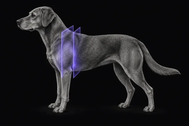
MRI of the canine brachial plexus for peripheral nerve sheath tumours
How MRI finds peripheral nerve sheath tumours of the canine brachial plexus, what the radiologist looks for, and what imaging can say about grade and extent.
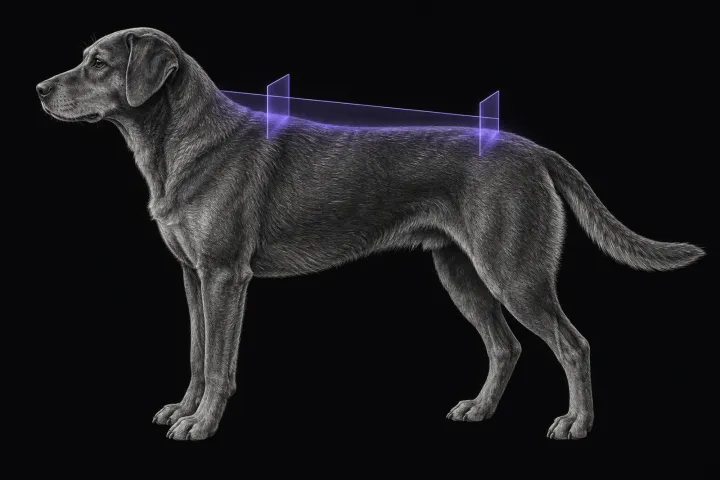
Spinal cord MRI for acute non-painful myelopathy in dogs
How MRI separates fibrocartilaginous embolism from acute non-compressive disc extrusion in dogs, when to scan, and which lesion measurements predict outcome.
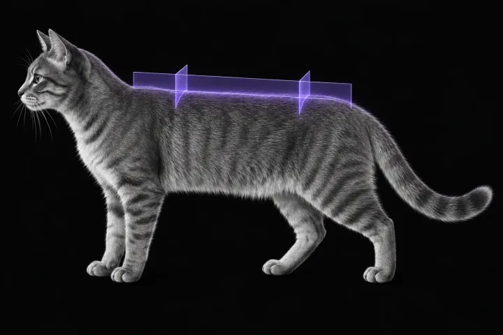
Spinal cord MRI in cats with myelopathy
How MRI separates lymphoma, FIP myelitis, ischaemic myelopathy and disc disease in cats with myelopathy, and where CSF and biopsy are still needed.