
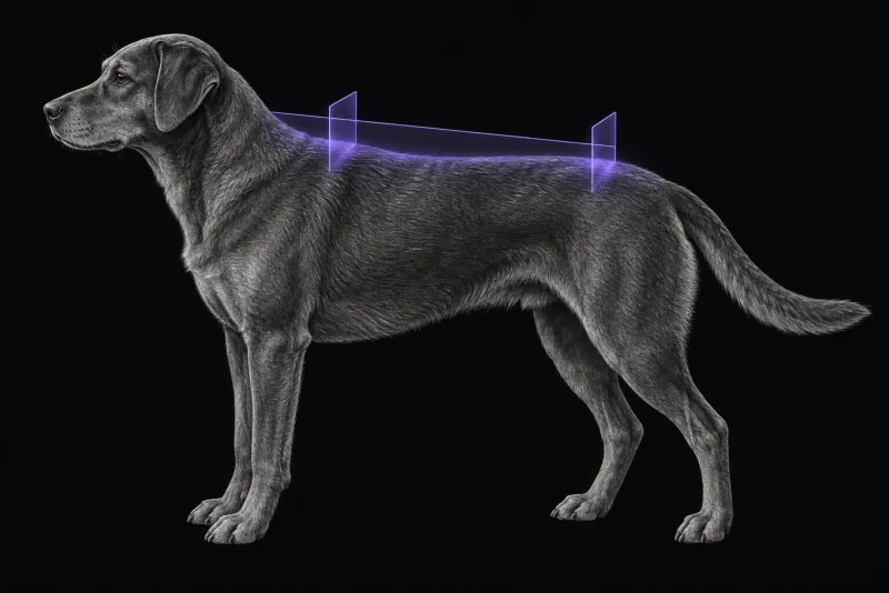
Two conditions that look the same at the door
Fibrocartilaginous embolic myelopathy (FCEM) and acute non-compressive nucleus pulposus extrusion (ANNPE) both cause a peracute onset (less than 6 hours) of spinal cord dysfunction that is generally non-progressive after the first 24–48 hours, according to a review of both conditions (De Risio, Frontiers in Veterinary Science, 2015). Onset during running, jumping or play is common in both, and so is vocalisation at onset.
Both can usually be told apart from compressive disc extrusion, fracture, myelitis and neoplasia. Telling them apart from each other is harder. Ante-mortem diagnosis rests largely on MRI, and treatment of both is conservative. In practice, MRI therefore does two jobs:
- it excludes a compressive or other surgical lesion
- it measures the cord lesion, which carries prognostic information in ischaemic myelopathy (De Risio et al., Journal of the American Veterinary Medical Association, 2008) and ANNPE (De Risio et al., Journal of the American Veterinary Medical Association, 2009)
Clinical clues before the scan
The review lists these clues:
- Age. FCEM occurs in immature and adult animals. ANNPE has been reported only in animals older than 1 year.
- Site. ANNPE most often affects the disc spaces between T12 and L2. FCEM has no such site predilection.
- Pain. Spinal hyperalgesia in the first days is uncommon in FCEM and was reported in 21–57% of dogs with ANNPE.
In 51 dogs with ischaemic myelopathy and 42 with ANNPE at one hospital (Fenn et al., Journal of the American Veterinary Medical Association, 2016), dogs with ANNPE were older and more likely to vocalise at onset, to have spinal hyperaesthesia, to have a C1–C5 lesion and to be ambulatory at discharge. Dogs with ischaemic myelopathy were more likely to have an L4–S3 lesion.
MRI features that separate the two
The patterns below come from the review unless another study is linked.
| Feature | FCEM | ANNPE |
|---|---|---|
| Position of the intramedullary T2 hyperintensity | Often over a vertebral body: 94% of lesions called ischaemic myelopathy were (Fenn et al., Veterinary Radiology & Ultrasound) | Over an intervertebral disc space: 85% of lesions called ANNPE were |
| Length on T2-weighted images | Generally longer than one vertebral length | Generally less than one vertebral length |
| Affected disc | Not part of the described pattern | Nucleus pulposus smaller with lower T2 signal; disc space often narrowed |
| Epidural space | Not part of the described pattern | Material or signal change dorsal to the disc in some dogs |
| Post-contrast T1 | Mild heterogeneous enhancement possible, generally on days 5–7 | Usually none; meningeal enhancement was associated with ANNPE in one study |
The FCEM lesion is typically focal, relatively sharply demarcated, often lateralised and mainly in the grey matter.
How reliable is the call?
- In 187 studies read by a neurologist and a radiologist, interobserver agreement was moderate (kappa 0.56), strongest for the lesion position shown in the table.
- In 201 dogs read by three board-certified observers (Mari et al., Veterinary Record, 2017), agreement was substantial to almost perfect (kappa 0.635–0.828).
- In a smaller study of 20 dogs (Specchi et al., Journal of the American Veterinary Medical Association, 2016), a non-longitudinal pattern of hyperintensity also favoured ANNPE and greater lesion length favoured ischaemic myelopathy.
Anecdotal reports suggest the distinction is harder with low-field MRI, suboptimal image definition and limited neuroimaging experience.
Spinal cord MRI protocol at a glance
The lesion position, length and cross-sectional area are judged on T2-weighted images in sagittal and transverse planes. Timing matters: MRI 24–72 hours after onset may show no intraparenchymal change in a dog with FCEM, and a repeat scan 72 hours or more after onset may reveal the lesion. Diffusion-weighted imaging has increased sensitivity for spinal cord infarction in the early stages, but is technically challenging in the spine.
What the radiologist looks at
Beyond the features in the table, the reader measures the lesion, because its size predicts outcome:
| Measure | Ischaemic myelopathy, 50 dogs | ANNPE, 42 dogs |
|---|---|---|
| Lesion length to vertebral length ratio (sagittal T2) | Above 2.0: 100% sensitivity for an unsuccessful outcome | Sagittal extent associated with outcome on univariable analysis |
| Lesion cross-sectional area on transverse T2 | 67% or more: 100% sensitivity for an unsuccessful outcome | Best single predictor of outcome on multivariable analysis |
- Specificity is modest. The review lists the specificity of the ischaemic myelopathy cut-offs as 62% and 52%, and, for ANNPE, a cross-sectional area of 90% or more as 86% sensitive and 96% specific for an unsuccessful outcome.
- Gradient echo signal. In the ANNPE series, intramedullary hypointensity on gradient echo images was also associated with outcome on univariable analysis, and 67% of dogs had a successful outcome.
- A third possibility. Intradural or intramedullary disc extrusion is rarer. A linear tract from the disc into the cord, hypointense on T2*-weighted gradient echo images, is highly suggestive of it.
What your report will include
Every Veheri case is read by a radiologist or neurologist, and when you order you can choose Diplomate-only reporting or the full specialist panel. Neurology reporting covers CT and MRI. You can chat directly with the specialist assigned to your case, and a simplified pet owner report helps you explain the findings to the owner.
When you take the report forward:
- The diagnosis is presumptive. The MRI patterns have not been validated against histopathology in large studies.
- An early scan with no cord change does not rule out FCEM. A repeat scan 72 hours or more after onset may reveal the lesion.
- Pain perception remains a strong prognostic factor. In paraplegic dogs with thoracolumbar FCEM or ANNPE (Togawa et al., Frontiers in Veterinary Science, 2024), with 26 having outcome data, 1 of 12 dogs without pain perception (8.3%) regained independent ambulation, against 9 of 14 with pain perception (64.3%).
- Talking to the owner: in 201 dogs with peracute non-progressive T3–L3 myelopathy, 99% regained ambulation and the presumptive diagnosis was not associated with motor recovery. Faecal incontinence was more frequent with ANNPE (23%) than FCEM (7.5%), but the single-hospital comparison, which included cervical and lumbosacral lesions, found long-term faecal incontinence more likely with ischaemic myelopathy. The two studies disagree, so discuss incontinence risk with care.
What to include when you request a spinal MRI report
- signalment: age, breed and sex
- the time of onset and the time from onset to scan
- activity or trauma at onset, and any vocalisation
- whether signs progressed after the first 24–48 hours
- neurolocalisation, lateralisation and spinal pain on examination
- pain perception in the affected limbs
How Veheri can help
Veheri works with veterinary radiologists and neurologists and reports spinal MRI studies remotely for clinics. For compressive disc disease, see MRI of the canine thoracolumbar spine, and for cats, spinal cord MRI in cats.
Sources
Each claim in this guide links to its source. The full citations:
- De Risio L. A review of fibrocartilaginous embolic myelopathy and different types of peracute non-compressive intervertebral disk extrusions in dogs and cats. Front Vet Sci. 2015;2:24. doi:10.3389/fvets.2015.00024
- De Risio L, Adams V, Dennis R, et al. Association of clinical and magnetic resonance imaging findings with outcome in dogs suspected to have ischemic myelopathy: 50 cases (2000-2006). J Am Vet Med Assoc. 2008;233(1):129-35. doi:10.2460/javma.233.1.129
- De Risio L, Adams V, Dennis R, McConnell FJ. Association of clinical and magnetic resonance imaging findings with outcome in dogs with presumptive acute noncompressive nucleus pulposus extrusion: 42 cases (2000-2007). J Am Vet Med Assoc. 2009;234(4):495-504. doi:10.2460/javma.234.4.495
- Fenn J, Drees R, Volk HA, De Decker S. Comparison of clinical signs and outcomes between dogs with presumptive ischemic myelopathy and dogs with acute noncompressive nucleus pulposus extrusion. J Am Vet Med Assoc. 2016;249(7):767-75. doi:10.2460/javma.249.7.767
- Fenn J, Drees R, Volk HA, De Decker S. Inter- and intraobserver agreement for diagnosing presumptive ischemic myelopathy and acute noncompressive nucleus pulposus extrusion in dogs using magnetic resonance imaging. Vet Radiol Ultrasound. 57(1):33-40 (published online 2015). doi:10.1111/vru.12289
- Specchi S, Johnson P, Beauchamp G, et al. Assessment of interobserver agreement and use of selected magnetic resonance imaging variables for differentiation of acute noncompressive nucleus pulposus extrusion and ischemic myelopathy in dogs. J Am Vet Med Assoc. 2016;248(9):1013-21. doi:10.2460/javma.248.9.1013
- Mari L, Behr S, Shea A, et al. Outcome comparison in dogs with a presumptive diagnosis of thoracolumbar fibrocartilaginous embolic myelopathy and acute non-compressive nucleus pulposus extrusion. Vet Rec. 2017;181(11):293. doi:10.1136/vr.104090
- Togawa G, Lewis MJ, Devathasan D. Outcome in paraplegic dogs with or without pain perception due to thoracolumbar fibrocartilaginous embolic myelopathy or acute non-compressive nucleus pulposus extrusion. Front Vet Sci. 2024;11:1406843. doi:10.3389/fvets.2024.1406843
Related reading
More imaging guides
All imaging guides
Spinal cord MRI in cats with myelopathy
How MRI separates lymphoma, FIP myelitis, ischaemic myelopathy and disc disease in cats with myelopathy, and where CSF and biopsy are still needed.
MRI of the canine brachial plexus for peripheral nerve sheath tumours
How MRI finds peripheral nerve sheath tumours of the canine brachial plexus, what the radiologist looks for, and what imaging can say about grade and extent.
Brain MRI for dogs with seizures
Which dogs with seizures need brain MRI, what the evidence says about diagnostic yield, what the radiologist looks for and what your report will include.