
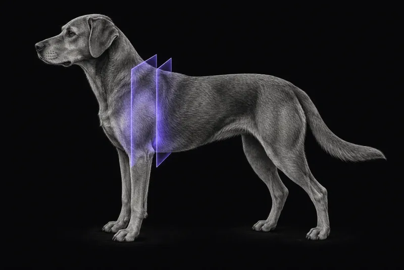
When thoracic limb lameness is neurological
Thoracic limb lameness is usually orthopaedic. As a review of neurological causes of thoracic limb lameness (Kerwin and Taylor, Veterinary Clinics of North America: Small Animal Practice, 2021) sets out, several neurological diseases also cause it:
- lateralised intervertebral disc extrusion
- caudal cervical spondylomyelopathy
- brachial plexus avulsion
- neuritis
- peripheral nerve sheath tumours (PNSTs)
Many of these compress or destroy the nerve roots of the cervical intumescence and produce a non-weight-bearing lameness, or root signature. Advanced imaging such as MRI is needed to find the cause.
Lameness from a nerve tumour is not always weakness. In one dog with a malignant tumour of the median nerve (Smith et al., Frontiers in Veterinary Science, 2025), the lameness resolved after resection, which the authors attribute to neuropathic pain rather than paresis.
Not every lesion in this region is a tumour. In another dog (Andruzzi et al., Journal of Veterinary Internal Medicine, 2021), a chronic brachial artery thrombus caused episodic non-weight-bearing lameness, limb atrophy, cool paw pads and a painful axilla. It mimicked a nerve sheath tumour and was confirmed with MRI, CT, ultrasound and surgery.
Where these tumours occur
- In 44 dogs with histologically confirmed spinal PNSTs (Morabito et al., Journal of Veterinary Internal Medicine, 2023), the brachial plexus (C6–T2) was the most common site (18 of 44). Median age was 9 years, and lameness was the most common reason for referral (20 of 44).
- In 30 dogs with presumptive or confirmed nerve sheath tumours (Cooper-Khan et al., Veterinary Sciences, 2024), 22 involved spinal nerves, mostly of the brachial plexus, and 13 of these invaded the spinal canal. Survival was generally longer without spinal canal or intracranial involvement.
- In nine dogs operated on for brachial plexus tumours extending into the vertebral canal (Potamopoulou et al., Veterinary Surgery, 2024), the eighth cervical nerve root was affected in seven.
Brachial plexus MRI protocol at a glance
Some brachial plexus tumours are hard to detect, so a study of 18 dogs with brachial plexus PNSTs (Kraft et al., Veterinary Radiology & Ultrasound, 2007) calls for a rigorous multiplanar, multi-sequence examination. Its key principles are transverse images that include both axillae and the vertebral canal in the same slice, so lesions show up as asymmetry, and contrast, which was critical for subtle disease and full extent. The 44-dog study used STIR first to find affected nerves, and noted its value on low-field scanners.
What the radiologist looks at
In the 18-dog study:
- 8 had diffuse thickening of one or more plexus nerves, extending into the vertebral canal in 6
- 10 had a nodule or mass in the axilla; seven of these also had diffuse nerve sheath thickening, which reached the vertebral canal in 3
- most tumours were hyperintense to muscle on T2-weighted images and isointense on T1-weighted images
- 8 enhanced only minimally to mildly, and 13 enhanced heterogeneously
In the 44-dog study, most tumours involved both the vertebral canal and the peripheral nerve, and only 8 had a dumbbell shape. Muscle atrophy was seen in 40 of 44. No tumour caused vertebral lysis, but 34 showed mild bony changes consistent with pressure atrophy.
The reader compares the two sides for asymmetry of nerves and muscles, follows the affected nerves towards the foramina and vertebral canal, and states how far the disease extends. Imaging is weaker on grade:
| MRI finding | Association with histological grade (44-dog study) |
|---|---|
| Large tumour volume | Grade 3 larger than grade 1 |
| Peripheral contrast enhancement | Most frequent in grade 3 (62.5%) |
| Homogeneous enhancement | Most frequent in grades 1 and 2 |
| Muscle atrophy, heterogeneous signal, growth into the vertebral canal | Not associated with grade |
What your report will include
Every Veheri case is read by a radiologist or neurologist, and when you order you can choose Diplomate-only reporting or the full specialist panel. Neurology reporting covers CT and MRI. You can chat directly with the specialist assigned to your case, and a simplified pet owner report helps you explain the findings to the owner.
When you take the report forward:
- Use the extent map for treatment planning. In 30 dogs with malignant PNSTs of the brachial or lumbosacral plexus treated surgically (Stokes et al., Veterinary and Comparative Oncology, 2023), 13 needed a laminectomy as well as amputation or hemipelvectomy. Margins were complete (R0) in 12, recurrence occurred in 13 (46%), and median disease-specific survival was 570 days.
- Expect recurrence to be a risk even with aggressive surgery. In the nine dogs treated with combined forequarter amputation and hemilaminectomy, four were euthanised for suspected recurrence.
- Grade needs histology. Grade was difficult to identify from imaging alone, and in the 30-dog series, 25 diagnoses were presumptive rather than histologically confirmed.
- Talking to the owner: spinal canal or intracranial involvement matters for outlook, because survival was generally longer without it.
What to include when you request a brachial plexus MRI report
- signalment: age, breed and sex
- duration and progression of the lameness, and whether the dog bears weight
- pain on palpation of the axilla or limb, and muscle atrophy
- neurological examination findings in the affected limb
- orthopaedic work-up already done and its results
- whether surgery or radiotherapy is being considered, so extent is reported in the detail treatment needs
How Veheri can help
Veheri provides veterinary teleradiology and teleneurology and reports MRI studies of the brachial plexus remotely for clinics. For other neurological presentations, see when veterinary MRI is the right move, and for a differential named above, MRI of the canine cervical spine for cervical spondylomyelopathy.
Sources
Each claim in this guide links to its source. The full citations:
- Kerwin SC, Taylor AR. Neurologic causes of thoracic limb lameness. Vet Clin North Am Small Anim Pract. 2021;51(2):357-364. doi:10.1016/j.cvsm.2020.12.003
- Smith J, Kent M, Glass E, Davis G. Case report: resolution of lameness via compartmental resection of a malignant nerve sheath neoplasm of the median nerve in a dog. Front Vet Sci. 2025;12:1551567. doi:10.3389/fvets.2025.1551567
- Andruzzi MN, Voges AK, Russell KE, Jeffery ND. Brachial artery thrombosis in a dog causing monoparesis mimicking nerve sheath tumor. J Vet Intern Med. 2021;35(5):2415-2420. doi:10.1111/jvim.16213
- Morabito S, Specchi S, Di Donato P, et al. Relationship between magnetic resonance imaging findings and histological grade in spinal peripheral nerve sheath tumors in dogs. J Vet Intern Med. 2023;37(6):2278-2290. doi:10.1111/jvim.16839
- Cooper-Khan RS, Frankovich AN, Thompson CA, et al. Clinical findings and outcome in 30 dogs with presumptive or confirmed nerve sheath tumors. Vet Sci. 2024;11(5):192. doi:10.3390/vetsci11050192
- Potamopoulou M, Petite A, Findji L. Combined forequarter amputation and hemilaminectomy for treatment of canine peripheral nerve sheath tumors of the brachial plexus invading the spinal canal: surgical technique and outcome in nine dogs. Vet Surg. 2024;53(8):1477-1484. doi:10.1111/vsu.14166
- Kraft S, Ehrhart EJ, Gall D, et al. Magnetic resonance imaging characteristics of peripheral nerve sheath tumors of the canine brachial plexus in 18 dogs. Vet Radiol Ultrasound. 2007;48(1):1-7. doi:10.1111/j.1740-8261.2007.00195.x
- Stokes R, Wustefeld-Janssens BG, Hinson W, et al. Surgical and oncologic outcomes in dogs with malignant peripheral nerve sheath tumours arising from the brachial or lumbosacral plexus. Vet Comp Oncol. 2023;21(4):739-747. doi:10.1111/vco.12938
Related reading
More imaging guides
All imaging guides
Brain MRI for dogs with seizures
Which dogs with seizures need brain MRI, what the evidence says about diagnostic yield, what the radiologist looks for and what your report will include.
MRI of the canine cervical spine for cervical spondylomyelopathy
How MRI characterises cervical spondylomyelopathy in dogs, what traction and flexion-extension studies add, and why compression alone does not prove the cause.
MRI of the craniocervical junction for Chiari-like malformation and syringomyelia
What MRI of the canine craniocervical junction should include for Chiari-like malformation and syringomyelia, and how to read a syrinx against the signs.