
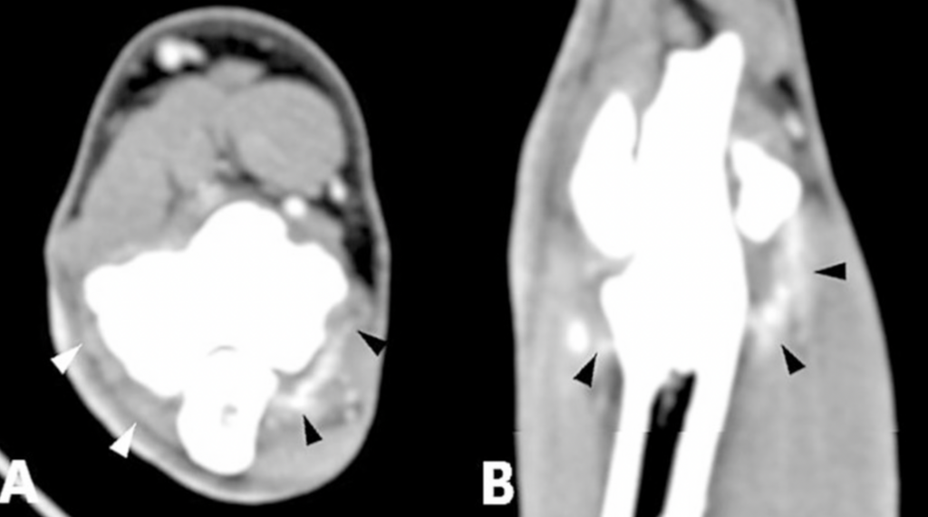
Is your dog still limping despite a “normal” CT scan? Learn how contrast-enhanced CT reveals hidden elbow inflammation and soft tissue injuries that standard scans miss, based on 2025 veterinary research.
Forelimb lameness is one of the most frustrating challenges for dog owners and veterinarians alike. While we often rely on standard X-rays or basic CT scans to find the culprit, a recent study from the Clinic for Small Animal Surgery at LMU Munich reveals that we might be missing the “soft” evidence.
By using intravenous contrast agents during a CT scan, specialists can now see internal inflammation and muscle damage that were previously invisible. Here is why this shift in imaging is becoming the new standard for canine orthopedics.
READ FULL ARTICLE: https://www.frontiersin.org/journals/veterinary-science/articles/10.3389/fvets.2025.1626472/full
The Problem with “Invisible” Elbow Pain
Standard CT scans are perfect for looking at bone—detecting fragments or cracks in the joint. However, many dogs suffer from Flexor Enthesopathy, a condition where the tendons and muscles attached to the elbow become inflamed and painful.
The LMU Munich study, which analyzed 326 elbow joints, found that a significant portion of these injuries don’t show up on a regular bone scan.
- The “Hidden” 32%: Nearly one-third of the dogs that showed muscle damage through contrast enhancement had no visible bone spurs or calcifications.
- Spotting Early Stages: Contrast agents act like a “highlighter,” revealing increased blood flow to injured tissues before the bone actually starts to change.
- Identifying Synovitis: The study showed that contrast-enhanced imaging is the best way to detect active inflammation in the joint capsule (synovitis), which often accompanies common issues like Medial Coronoid Process Disease (MCPD).
Understanding the Results: Contrast vs. Standard Imaging
The research team tracked 605 different pathological findings to see how contrast changed the diagnosis. The results were clear: contrast provides a level of detail that “dry” CT simply cannot.
| Feature | What Standard CT Sees | What Contrast CT Reveals |
|---|---|---|
| Bone Fragments | Visible as loose pieces. | Shows if the surrounding tissue is actively inflamed. |
| Arthritis | Detects bone spurs (osteophytes). | Shows the severity of the joint’s inflammatory response. |
| Muscle Tears | Hard to distinguish from healthy tissue. | Highlights damaged areas with hyper-attenuated “rims”. |
| Early Enthesopathy | Often missed entirely. | Identifies soft tissue changes before bone spurs form. |
Is Contrast Safe for Your Dog?
While the diagnostic benefits are high, the study was transparent about the risks. Out of 163 dogs, only one (0.8%) had a serious adverse reaction involving heart rhythm. While this is a very low percentage, the researchers emphasize that vets should always check a dog’s history and kidney health before proceeding.
The Verdict: Should Your Dog Get a Contrast CT?
The study concludes that a contrast agent should be administered during most elbow CT scans to ensure no “invisible” pathological changes are overlooked. This is especially true if:
- The dog is lame but standard X-rays look normal.
- The vet suspects Flexor Enthesopathy.
- You need to distinguish between an old injury and an active, painful one.
Conclusion
The study conducted at LMU Munich makes a compelling case for the routine use of contrast agents in canine elbow imaging. While standard CT remains a powerful tool for identifying bony fragments, it often fails to capture the full scope of soft tissue inflammation and early-stage muscle disease. By utilizing contrast-enhanced CT, veterinarians can identify “invisible” conditions like flexor enthesopathy in dogs that show no structural bone changes on regular scans. Ultimately, this extra step allows for a more accurate diagnosis, a better-defined prognosis, and a treatment plan that addresses the true source of a dog’s pain. However, because a small risk of adverse reactions (0.8%) exists, the decision should always be tailored to the individual patient’s health profile.
Start today: Book your DEMO Smarter Teleradiology. Faster Diagnosis. Better Results.
Imaging guides on this topic
Browse all imaging guides
CT of the canine elbow for medial coronoid disease
When CT is worth it for medial coronoid disease in dogs, what it finds that radiographs miss, and where arthroscopy still gives the better answer.
MRI of the canine shoulder for biceps and supraspinatus tendinopathy
What MRI shows in canine biceps and supraspinatus tendinopathy, how it compares with CT arthrography, and why a bright tendon alone is not disease.
MRI of the canine stifle for cruciate and meniscal injury
How accurately MRI shows cruciate rupture and meniscal tears in dogs, how field strength and implants affect it, and where arthroscopy still matters.