
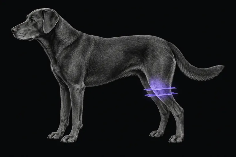
Why image the stifle with MRI
Cranial cruciate ligament (CCL) rupture in dogs is often bilateral and asymmetrical (Fazio et al., Veterinary Radiology & Ultrasound), and ranges from partial to complete rupture. Damage to other structures in the joint can occur alongside it, and clinical detection of that damage is unreliable (Mattei et al., Veterinary Radiology & Ultrasound, 2025). Inspecting the joint during surgery is advised, but it adds to the risk of complications. MRI offers a non-invasive alternative that also shows the soft tissues around the joint.
MRI is most useful when it answers a question that changes the plan:
- is a palpably stable stifle a partial tear?
- is the meniscus torn?
- why is a dog lame again after surgery?
Partial or complete cruciate rupture
- 3 T, 28 dogs (56 stifles). Each dog had complete rupture on one side and partial rupture on the other. Against arthroscopy, fibre loss on MRI had a sensitivity of 0.78 and a specificity of 0.50–0.60 for complete rupture, and a specificity of 1.00 for partial tears. A visual analogue scale score of 79 or more indicated complete rupture. MRI was specific in stable partial rupture and sensitive but not specific in unstable complete rupture.
- Single high-field sequence, 21 stifles (18 compared with surgery). A single sagittal sequence separated surgically confirmed complete from partial rupture with a sensitivity of 85.7% and a specificity of 75%.
- 3 T short protocol, 21 dogs. In a study of non-traumatic CCL rupture (Galindo-Zamora et al., BMC Veterinary Research, 2013), all cranial cruciate ligaments were correctly identified as ruptured, and all but one caudal cruciate ligament as intact.
Meniscal tears: accuracy depends on scanner and reader
| Study | Result for meniscal tears |
|---|---|
| 0.5 T, 42 stifles (Böttcher et al., Veterinary Surgery, 2010) | Sensitivity 0.64 and specificity 0.90 against arthroscopy. 14 of 22 tears needing meniscectomy were identified. |
| Low-field, 50 stifles, 15 observers (Böttcher et al., Veterinary Radiology & Ultrasound, 2012) | Medial meniscus: median sensitivity 0.74, specificity 0.89. Lateral meniscus: median sensitivity 0.00. Interrater agreement fair. |
| 0.25 T, 57 stifles (Knebel et al., Animals, 2024) | Agreement between a surgeon and a radiologist was high (κ = 0.86), and MRI findings correlated significantly with findings at surgery. |
| 31 stifles, medial meniscus (Franklin et al., Journal of the American Veterinary Medical Association, 2017) | MRI sensitivity 75% and specificity 100%; ultrasonography sensitivity 95% and specificity 82% (unblinded surgeon as reference). |
| 3 T short protocol, 21 dogs | High sensitivity and specificity, with occasional misdiagnosis. |
| Single high-field sequence | 7 of 8 surgically confirmed injured menisci identified. |
Two messages stand out. The authors of the 0.5 T study warn that a negative low-field MRI should be interpreted with caution, because many tears were missed. And accuracy in the 15-observer study was observer-dependent, with no clear link to experience.
After stabilisation surgery
When lameness returns after surgery, a late meniscal tear is one question MRI can address. In 8 stifles from large breed dogs imaged after tibial tuberosity advancement, or TTA (Taylor-Brown et al., Veterinary and Comparative Orthopaedics and Traumatology, 2014), MRI identified a medial meniscal tear in 6, all confirmed at surgery. In the other 2, the menisci looked intact and no tear was found. Implant artefacts were present but did not prevent interpretation.
Implant material matters. In cadaver limbs at 1.5 T (David et al., Veterinary Radiology & Ultrasound, 2012), a stainless steel tibial plateau levelling osteotomy (TPLO) implant obscured or distorted most of the stifle anatomy. Titanium TTA implants and the steel crimps of an extracapsular stabilisation mainly affected the lateral stifle, and the cruciate ligaments and medial meniscus could still be assessed.
Stifle MRI protocol at a glance
Stifle MRI has been studied on scanners from 0.25 T to 3 T, and meniscal accuracy depends on both the scanner and the reader. At high field, even a single sagittal sequence separated complete from partial rupture and measured the tibial plateau angle. With implants in place, turbo spin-echo sequences without fat saturation showed less artefact.
What the radiologist looks at
- The ligament. Fibre loss and the overall appearance of the CCL, read against arthroscopy-tested criteria for partial and complete rupture.
- Both menisci. The lateral meniscus is the harder one: in the 15-observer study, median sensitivity was 0.00.
- More than the ligament. The single-sequence study also found abnormal subchondral bone signal in nine stifles and muscle hyperintensity in six. The authors suggest these findings may have prognostic value.
- Planning measurements. Tibial plateau angle measured on that sequence correlated strongly with radiographic measurement (Pearson coefficient 0.923).
- What it cannot judge well. With the 3 T short protocol, cartilage damage and the degree of osteoarthritis could not be properly evaluated.
What your report will include
Every Veheri case is read by a radiologist or neurologist, and when you order you can choose Diplomate-only reporting or the full specialist panel. You can chat directly with the specialist assigned to your case, and a simplified pet owner report helps you explain the findings to the owner.
When you take the report forward:
- A negative MRI with high suspicion. When MRI is negative but suspicion of a meniscal tear stays high, joint inspection is still worth discussing.
- Lameness after surgery. MRI can look for a late meniscal tear, but the implant type decides how much of the joint can be assessed.
- The other stifle. Disease is often bilateral, so keep the opposite joint in mind.
What to include when you request a stifle MRI report
- signalment: age, breed and sex
- which limb, duration of lameness, and drawer or tibial thrust findings
- the state of the other stifle, since disease is often bilateral
- previous surgery, the technique used and the implant material
- recent radiographs
- the exact question: partial versus complete tear, meniscal status, or lameness after surgery
How Veheri can help
Veheri’s radiologists report veterinary MRI and CT studies remotely for clinics. For other lame dogs, see MRI of the canine shoulder and contrast-enhanced CT for limping dogs.
Sources
Each claim in this guide links to its source. The full citations:
- Fazio CG, Muir P, Schaefer SL, Waller KR. Accuracy of 3 Tesla magnetic resonance imaging using detection of fiber loss and a visual analog scale for diagnosing partial and complete cranial cruciate ligament ruptures in dogs. Vet Radiol Ultrasound. 59(1):64-78 (published online 2017). doi:10.1111/vru.12567
- Mattei C, Pratesi A, Bernardini M, Specchi S. May a single presurgical high-field MRI sequence replace standard radiographs for TPLO surgical planning in dogs? Vet Radiol Ultrasound. 2025;66(1):e70005. doi:10.1111/vru.70005
- Galindo-Zamora V, Dziallas P, Ludwig DC, et al. Diagnostic accuracy of a short-duration 3 Tesla magnetic resonance protocol for diagnosing stifle joint lesions in dogs with non-traumatic cranial cruciate ligament rupture. BMC Vet Res. 2013;9:40. doi:10.1186/1746-6148-9-40
- Böttcher P, Brühschwein A, Winkels P, et al. Value of low-field magnetic resonance imaging in diagnosing meniscal tears in the canine stifle: a prospective study evaluating sensitivity and specificity in naturally occurring cranial cruciate ligament deficiency with arthroscopy as the gold standard. Vet Surg. 2010;39(3):296-305. doi:10.1111/j.1532-950X.2010.00650.x
- Böttcher P, Armbrust L, Blond L, et al. Effects of observer on the diagnostic accuracy of low-field MRI for detecting canine meniscal tears. Vet Radiol Ultrasound. 2012;53(6):628-35. doi:10.1111/j.1740-8261.2012.01967.x
- Knebel J, Wilke SK, Neumann S, et al. Low-field magnetic resonance imaging for the detection of medial meniscal lesions in cranial cruciate-deficient stifle joints in dogs. Animals (Basel). 2024;14(21):3097. doi:10.3390/ani14213097
- Franklin SP, Cook JL, Cook CR, et al. Comparison of ultrasonography and magnetic resonance imaging to arthroscopy for diagnosing medial meniscal lesions in dogs with cranial cruciate ligament deficiency. J Am Vet Med Assoc. 2017;251(1):71-79. doi:10.2460/javma.251.1.71
- Taylor-Brown F, Lamb CR, Tivers MS, Li A. Magnetic resonance imaging for detection of late meniscal tears in dogs following tibial tuberosity advancement for treatment of cranial cruciate ligament injury. Vet Comp Orthop Traumatol. 2014;27(2):141-6. doi:10.3415/VCOT-13-08-0107
- David FH, Grierson J, Lamb CR. Effects of surgical implants on high-field magnetic resonance images of the normal canine stifle. Vet Radiol Ultrasound. 2012;53(3):280-8. doi:10.1111/j.1740-8261.2011.01901.x
Related reading
More imaging guides
All imaging guides
MRI of the canine brachial plexus for peripheral nerve sheath tumours
How MRI finds peripheral nerve sheath tumours of the canine brachial plexus, what the radiologist looks for, and what imaging can say about grade and extent.
Brain MRI for dogs with seizures
Which dogs with seizures need brain MRI, what the evidence says about diagnostic yield, what the radiologist looks for and what your report will include.
MRI of the canine cervical spine for cervical spondylomyelopathy
How MRI characterises cervical spondylomyelopathy in dogs, what traction and flexion-extension studies add, and why compression alone does not prove the cause.