
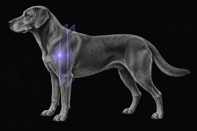
Why shoulder soft-tissue disease needs more than radiographs
Supraspinatus tendinopathy occurs in a mineralised and a non-mineralised form. In 24 dogs treated surgically (Lafuente et al., Veterinary Surgery, 2009), the most consistent findings were:
- chronic, unilateral lameness
- lameness that was intermittent or waxing and waning
- pain on palpation of the cranial shoulder
The non-mineralised form has no mineral for a radiograph to show, and the authors describe ultrasonography and MRI as good techniques for detecting it.
The biceps and supraspinatus tendons are close neighbours at the front of the shoulder, and an enlarged supraspinatus tendon can displace the biceps tendon. On ultrasonography or MRI of 35 shoulders in the same study, the supraspinatus tendon was enlarged in 54% and had increased fluid content in 63%. The biceps tendon was displaced medially in 60%.
How well MRI matches surgical findings
In 21 dogs with soft-tissue shoulder disease confirmed at arthroscopy or open surgery (Murphy et al., Veterinary Surgery, 2008), MRI was compared structure by structure. Agreement meant MRI and surgery agreed on normal versus abnormal. Concordance meant MRI named the same lesion type as surgery.
| Structure | Agreement | Concordance |
|---|---|---|
| Biceps tendon | 90% | 100% |
| Biceps tendon impingement | 90% | 100% |
| Subscapularis | 95% | 62% |
| Medial glenohumeral ligament | 84% | 83% |
| Lateral glenohumeral ligament | 88% | 100% |
| Infraspinatus | 100% | 100% |
What supraspinatus tendinopathy looks like on MRI
- Tendon size. In dogs with surgically treated, histologically confirmed non-mineralised tendinopathy (Spall et al., Veterinary Surgery, 2016), median supraspinatus tendon volume on MRI was 1,323 mm³, against 630 mm³ in dogs with other orthopaedic disease and 512 mm³ in healthy dogs.
- Effects on the biceps. In 27 dogs with non-mineralised tendinopathy (Piras et al., Animals, 2023), MRI showed a distended biceps tendon sheath in 10, compression of the biceps tendon sheath in 5 and supraspinatus enlargement in 3. Histology showed severe myxomatous degeneration in all 27.
- Both sides. Of 12 dogs with bilateral imaging abnormalities, 11 were lame on one side only.
The pitfall: a normal tendon can look abnormal
In non-lame Beagles (Pownder et al., American Journal of Veterinary Research, 2018), every supraspinatus tendon had a three-layer appearance on MRI: a thick, hyperintense central layer between thin hypointense margins. Histology showed the central layer was water-rich glycosaminoglycan with irregular collagen. The authors conclude that tendinosis should not be diagnosed on hyperintense signal alone; other MRI evidence of shoulder disease and other diagnostic findings are needed.
Ultrasound shows the same trap: a central hypoechoic area near the insertion was present in all normal tendons examined (Lassaigne et al., Veterinary Record) and should be considered normal without other criteria.
MRI compared with CT arthrography and arthroscopy
- for the biceps tendon and sheath, CTA had a sensitivity of 71% and specificity of 75%
- it was of limited value for the medial and lateral glenohumeral ligaments (sensitivity 13% and 0%) and the subscapularis tendon (sensitivity 14%)
- CT and CTA did add information on osteophytes, subchondral defects and joint mice
The authors conclude CTA is an adjunct, not a replacement for arthroscopy. These CT arthrography figures and the MRI figures above come from different small retrospective studies with different reference standards, so they should not be compared directly.
Shoulder MRI protocol at a glance
The shoulder tendons and ligaments are seen best in different planes, so the study combines several. A study of scan plane, positioning and arthrography (Agnello et al., Veterinary Radiology & Ultrasound, 2008) found that limb extension improved visibility of the medial structures, and that MR arthrography with the joint extended gave the best visualisation of all structures.
What the radiologist looks at
- Tendon size. An enlarged supraspinatus tendon, and tendon volume, which separated tendinopathy from other disease.
- The biceps tendon and sheath. Distension, compression or medial displacement by the supraspinatus.
- Signal, read with care. A hyperintense central layer can be normal tendon, so signal is weighed with size, the biceps sheath and the clinical findings.
- The other shoulder. Changes are often bilateral, so a finding is most convincing when it sits in the painful shoulder and fits the examination.
What your report will include
Every Veheri case is read by a radiologist or neurologist, and when you order you can choose Diplomate-only reporting or the full specialist panel. You can chat directly with the specialist assigned to your case, and a simplified pet owner report helps you explain the findings to the owner.
When you take the report forward:
- Match the findings to the examination. A bright tendon alone is not proof of tendinosis, and bilateral changes often come with lameness on one side only.
- CT arthrography does not replace arthroscopy. It is an adjunct that adds information on osteophytes, subchondral defects and joint mice.
- Lesion type in the subscapularis is less certain. Concordance with surgery was 62% on MRI, against 100% for the biceps tendon.
- Talking to the owner: imaging changes in the other shoulder do not always mean that shoulder is causing the lameness.
What to include when you request a shoulder MRI report
- signalment: age, breed and sex
- which limb, duration and pattern of lameness
- where pain is elicited on examination, for example over the biceps tendon or the supraspinatus insertion
- radiographs, including any mineralisation near the greater tubercle
- whether both shoulders should be imaged, given how often changes are bilateral
- previous treatment, injections or surgery
How Veheri can help
Veheri’s radiologists report veterinary MRI and CT studies remotely for clinics, and can support your team with MRI scanning protocols and positioning for the shoulder. For related reading, see MRI of the canine stifle and why contrast-enhanced CT helps limping dogs.
Sources
Each claim in this guide links to its source. The full citations:
- Lafuente MP, Fransson BA, Lincoln JD, et al. Surgical treatment of mineralized and nonmineralized supraspinatus tendinopathy in twenty-four dogs. Vet Surg. 2009;38(3):380-7. doi:10.1111/j.1532-950X.2009.00512.x
- Piras LA, Olimpo M, Lafuente P, et al. Surgical treatment of nonmineralized supraspinatus tendinopathy in dogs: a retrospective long-term follow-up. Animals (Basel). 2023;13(4):592. doi:10.3390/ani13040592
- Murphy SE, Ballegeer EA, Forrest LJ, Schaefer SL. Magnetic resonance imaging findings in dogs with confirmed shoulder pathology. Vet Surg. 2008;37(7):631-8. doi:10.1111/j.1532-950X.2008.00429.x
- Spall BF, Fransson BA, Martinez SA, Wilkinson TE. Tendon volume determination on magnetic resonance imaging of supraspinatus tendinopathy. Vet Surg. 2016;45(3):386-91. doi:10.1111/vsu.12452
- Pownder SL, Caserto BG, Hayashi K, et al. Magnetic resonance imaging and histologic features of the supraspinatus tendon in nonlame dogs. Am J Vet Res. 2018;79(8):836-844. doi:10.2460/ajvr.79.8.836
- Lassaigne CC, Boyer C, Sautier L, Taeymans O. Ultrasound of the normal canine supraspinatus tendon: comparison with gross anatomy and histology. Vet Rec. 186(17):e14 (published online 2019). doi:10.1136/vr.105552
- Agnello KA, Puchalski SM, Wisner ER, et al. Effect of positioning, scan plane, and arthrography on visibility of periarticular canine shoulder soft tissue structures on magnetic resonance images. Vet Radiol Ultrasound. 2008;49(6):529-39. doi:10.1111/j.1740-8261.2008.00429.x
- Eivers CR, Corzo-Menéndez N, Austwick SH, et al. Computed tomographic arthrography is a useful adjunct to survey computed tomography and arthroscopic evaluation of the canine shoulder joint. Vet Radiol Ultrasound. 2018;59(5):535-544. doi:10.1111/vru.12670
Related reading
More imaging guides
All imaging guides
MRI of the canine brachial plexus for peripheral nerve sheath tumours
How MRI finds peripheral nerve sheath tumours of the canine brachial plexus, what the radiologist looks for, and what imaging can say about grade and extent.
Brain MRI for dogs with seizures
Which dogs with seizures need brain MRI, what the evidence says about diagnostic yield, what the radiologist looks for and what your report will include.
MRI of the canine cervical spine for cervical spondylomyelopathy
How MRI characterises cervical spondylomyelopathy in dogs, what traction and flexion-extension studies add, and why compression alone does not prove the cause.