
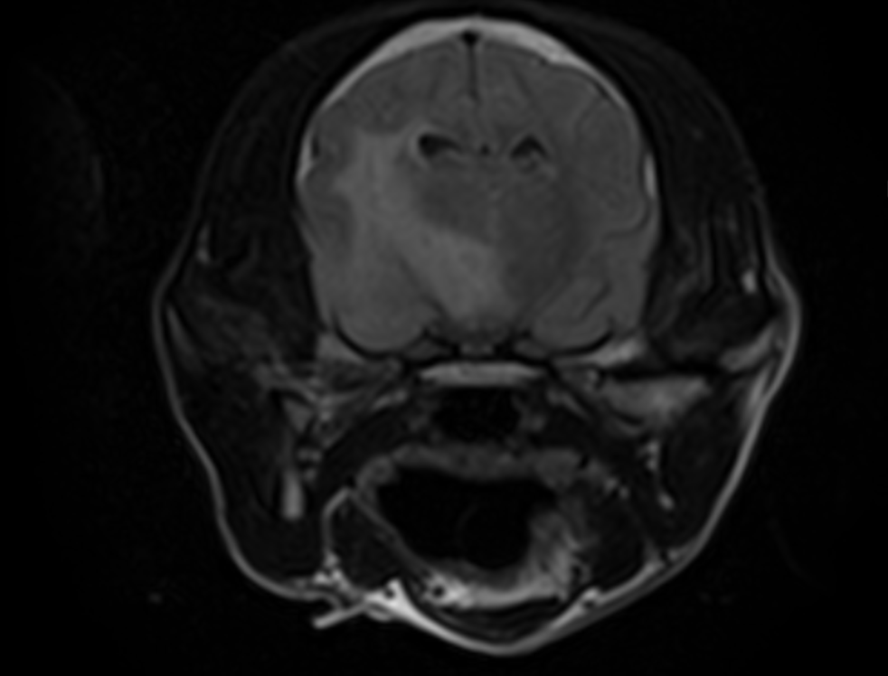
Summary MRI is central to canine neurological imaging because it answers the hardest clinical questions about brain and spinal cord disease. Following consensus protocols, coordinating anesthesia, and using advanced sequences when needed will materially improve diagnostic confidence and care decisions.
If you’re deciding whether a dog needs advanced brain or spine imaging, here’s the bottom line: MRI is the leading modality for soft-tissue detail in canine neurology, guiding diagnosis and treatment plans with the highest confidence.
What canine neurological imaging covers (and why MRI leads)
Canine neurological imaging spans radiography, CT, MRI, and occasionally ultrasound for peripheral structures. For intracranial and spinal cord disease, MRI’s superior soft-tissue contrast makes it the default choice. Clinicians use it to evaluate seizures, mentation changes, vestibular signs, acute paralysis, neck/back pain, trauma, and suspected neoplasia or inflammation.
- Typical indications: refractory seizures, suspected IVDD, brain tumors, encephalitis/meningitis, stroke-like events, cranial nerve deficits.
- MRI advantages: multiplanar acquisition; tissue contrast for gray/white matter; edema, hemorrhage, inflammation and neoplasia characterization; contrast enhancement patterns.
“A 6–7 sequence epilepsy-specific MRI protocol for veterinary patients is proposed and further advanced MR and functional imaging is reviewed.” — Clare Rusbridge, BMC Veterinary Research
Core veterinary MRI sequences and protocols
High-field protocols typically include T2-weighted, T2-FLAIR, T1-weighted pre- and post-gadolinium, diffusion-weighted imaging (where available), and susceptibility-sensitive sequences.
Field strength influences choices; high-field systems better detect hemorrhage and subtle hippocampal pathologies. For orientation and neuroanatomy refreshers, the UMN canine MRI atlas is a practical reference. For inflammatory CNS context in dogs, see: https://www.merckvetmanual.com/nervous-system/meningitis-encephalitis-and-encephalomyelitis/meningitis-encephalitis-and-encephalomyelitis-in-animals.
“T2W, T2W FLAIR and T1W images pre and post paramagnetic contrast (gadolinium based) are included as standard in most protocols used by specialists who are active in the veterinary field.” — Clare Rusbridge, BMC Veterinary Research
When to choose MRI versus CT
CT excels at bone, trauma triage, and surgical planning for osseous lesions; MRI excels at brain, spinal cord, meninges, intervertebral discs, and soft tissue. Practical decision-making balances the suspected pathology, anesthesia risk, and local availability. For intracranial neoplasia, MRI is generally preferred: https://www.merckvetmanual.com/nervous-system/neoplasia-of-the-nervous-system/neoplasia-of-the-nervous-system-in-small-animals. A case study from RVC shows MRI clarifying spinal cord injury while CT identified a vertebral fracture, underlining modality complementarity.
Anesthesia, safety, and client communication
Most dogs require general anesthesia for diagnostic-quality MRI. Close collaboration with anesthesia teams is essential for patient stability and intracranial pressure management, particularly in suspected mass lesions or herniation risk. Set expectations with owners on fasting, monitoring, recovery, and how results directly influence treatment, surgery, and prognosis.
Cornell’s Neurology & Neurosurgery service illustrates this coordination and impact.
“General anesthesia is sometimes needed in order to stop the seizures and also allows for a brain MRI and cerebrospinal fluid collection to look for any cause(s) that could potentially elicit the seizure events.” — Cornell University Hospital for Animals, Neurology & Neurosurgery
Advanced MRI: when to go beyond “basic” sequences
When conventional MRI is non-diagnostic or equivocal, advanced techniques can improve detection and characterization. In epilepsy cases, volumetrics and higher-resolution hippocampal imaging refine phenotypes and research insights. For neoplasia, dynamic contrast-enhanced MRI (DCE-MRI) may differentiate meningiomas from other tumors. For infectious/inflammatory disease, consistent sequence combinations and pattern recognition improve differential diagnostics, including protozoal diseases like neosporosis.
“Three main aims of advanced MRI in the epileptic animal are: (1) to rule out causes of epileptic seizures which may be treatable with means other than antiseizure medications (ASM) only (e.g., inflammatory or infectious brain disease), (2) to identify lesions caused by epileptic seizures which are not themselves the source of seizures, and (3) to provide data to further advance the field of research into the pathogenesis and/or treatment of epilepsy (5).” — Kari. D. FossFrontiers in Veterinary Science
“In veterinary medicine MRI is routinely used to identify and classify canine brain tumours, being the preferred modality to assess for intracranial disease” — Sarah Cussen et al., Frontiers in Veterinary Science
Reading MRI findings: common patterns clinicians watch for
Every case is different, but pattern recognition helps. Inflammatory disease often shows multifocal T2/FLAIR hyperintensities and possible meningeal enhancement (overview above via Merck). Neoplasia may present with mass effect, enhancement, and perilesional edema (Merck neoplasia overview linked above). Infectious protozoal disease (neosporosis) can show T2/FLAIR hyperintense rims and T1 hypointensity with variable enhancement.
Suggested figure: Core brain MRI sequences by indication
- Data: Indications (epilepsy, neoplasia, inflammatory/infectious, acute stroke-like, trauma) mapped to core sequences (T2, FLAIR, T1 pre/post, DWI, GRE/SWI), with “recommended” vs “optional based on field strength.”
- Format: Simple table with checkmarks for core vs optional.
- Alt text: “Matrix showing recommended MRI sequences for common canine neurologic indications.”
Smarter Teleradiology. Faster Diagnosis. Better Results Join the Veheri Community! Start today: Book your DEMO
Imaging guides on this topic
Browse all imaging guides
Brain CT in dogs when MRI is not available
What brain CT can and cannot detect in dogs when MRI is not available, for masses, stroke, inflammation and head trauma, and when MRI or CSF is still needed.
MRI of the craniocervical junction for Chiari-like malformation and syringomyelia
What MRI of the canine craniocervical junction should include for Chiari-like malformation and syringomyelia, and how to read a syrinx against the signs.
MRI of the canine thoracolumbar spine for disc extrusion
What MRI shows in dogs with acute thoracolumbar disc extrusion, how it compares with CT, and which findings matter for surgery, prognosis and myelomalacia.