
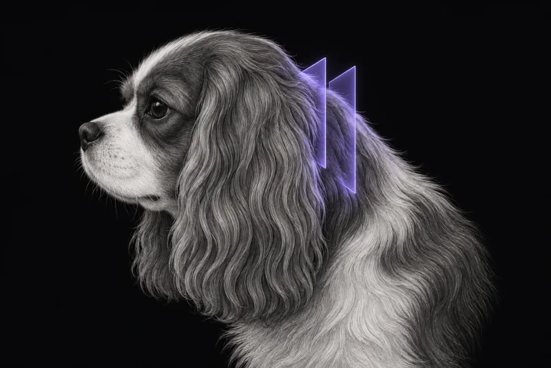
Why an abnormal scan is not always the answer
Chiari-like malformation (CM) is a complex disorder of the skull and craniocervical junction, associated with brachycephaly, a short skull base and crowding of the brain and cervical spinal cord. As a review of imaging in CM and SM (Rusbridge et al., Frontiers in Veterinary Science, 2018) explains, it is one of the most common causes of syringomyelia (SM): fluid-filled cavities in the spinal cord that develop when cerebrospinal fluid (CSF) channels are disrupted.
Finding a herniation or a syrinx on MRI is the easy part. Deciding whether they explain the signs is harder. In the Cavalier King Charles spaniel (CKCS), CM as defined by the BVA/KC health scheme is ubiquitous, and SM is common but can be asymptomatic.
- Screened dogs without signs. In 386 Australian CKCS screened between 2008 and 2024 (McLeay and Child, Australian Veterinary Journal), SM (a cavity of 1.0 mm or more) was found in 50.5%. Prevalence rose with age and did not fall over the 16 years.
- Other breeds. In 53 Chihuahuas (Kiviranta et al., Journal of Veterinary Internal Medicine, 2017), CM was present in 100% and SM in 38%. Many dogs had similar clinical signs without SM.
- Syrinxes change. In Pomeranians scanned twice (Jansma et al., Veterinary Sciences, 2025), 39.6% had developed SM or shown substantial progression by the second MRI, and 20.7% of dogs first classified as free of SM had developed it.
Signs that point to CM pain or symptomatic SM
In 130 CKCS with MRI of the neuroaxis and a diagnosis of CM pain or SM (Rusbridge et al., Journal of Veterinary Internal Medicine, 2019), the commonest signs were:
- vocalisation (65.4%)
- spinal pain (54.6%)
- reduced activity (37.7%)
- reduced ability to jump or use stairs (35.4%)
- touch aversion (30.0%)
- altered emotional state (28.5%)
- sleep disturbance (22%)
Signs of pain did not depend on SM. Phantom scratching, scoliosis, weakness and postural deficits were seen only in dogs with a syrinx 4 mm wide or more.
Phantom scratching was associated with a large dorsolateral syrinx (Nalborczyk et al., BMC Veterinary Research, 2017) reaching the superficial dorsal horn in the C3–C6 spinal cord segments (C2–C5 vertebrae). If a scratching dog with SM has no such syrinx, the authors advise looking for other causes of the scratching.
How far the syrinx extends
Extent matters. In CKCS, 76% of dogs with a syrinx at C1–C4 also had one in the C5–T1 and T2–L2 regions. In 18 dogs with holocord syringomyelia (Douralidou et al., Frontiers in Veterinary Science), the syrinx reached L3 to L6 caudally, and 17 had Chiari-like and/or other intracranial or craniocervical junction malformations.
Craniocervical junction MRI protocol at a glance
For a dog with signs, the 2018 imaging review recommends imaging the brain, craniocervical junction and the cervical and thoracic spinal cord, with T1- and T2-weighted images and transverse images through the widest part of any syrinx. The lumbar spine, contrast and additional sequences are added when the signs or the first images call for them. The limited three-sequence protocol offered to some breeders shows neither the brain nor the thoracolumbar cord, and is not recommended for a dog with clinical signs.
What the radiologist looks at
The findings are weighed against the signs you report. The links below follow the 2018 imaging review:
| MRI finding | What the evidence links it to |
|---|---|
| Rostral forebrain flattening, increased cranial height, loss of sulci definition, ventriculomegaly, reduced cisterna magna | Supports CM pain, together with consistent signs and exclusion of other causes |
| Rostral displacement of atlas and axis, increased odontoid angulation, medullary elevation | Craniocervical junction changes more likely in dogs with SM |
| Syrinx 4 mm wide or more | Signs of myelopathy in CKCS (Rusbridge et al., 2019) |
| Syrinx reaching the superficial dorsal horn mid-cervically | Phantom scratching and cervicotorticollis (Nalborczyk et al.) |
| Fluid signal void in the syrinx, expansive asymmetrical shape | An “active” filling syrinx. A quiescent syrinx is central and symmetrical |
- Head position. Cerebellar herniation is significantly greater with the head flexed than in the usual extended position.
- Signs SM does not explain. Seizures, facial nerve paralysis or generalised pruritus are not explained by SM, which is a spinal cord disease.
- Other causes. Cystic intramedullary tumours must be distinguished from SM, and contrast helps when there is a mass or the cause of CSF obstruction is not apparent.
What your report will include
Every Veheri case is read by a radiologist or neurologist, and when you order you can choose Diplomate-only reporting or the full specialist panel. Neurology reporting covers CT and MRI. You can chat directly with the specialist assigned to your case, and a simplified pet owner report helps you explain the findings to the owner.
When you take the report forward:
- CM pain is a diagnosis of exclusion. There is no objective MRI measure of CM pain; it rests on the weight of clinical and MRI evidence.
- Scratching without a matching syrinx is a reason to look for other causes.
- A previous scan helps. Syrinxes can develop or progress between scans, so comparing syrinx width over time is useful.
- Other modalities. Radiographs are not recommended for the investigation. CT has limited value beyond confirming herniation and defining bony craniocervical abnormalities, but should be performed when a vertebral malformation may need surgical fixation.
- Talking to the owner: SM is common and can be asymptomatic, so a syrinx on the scan does not by itself explain every sign.
What to include when you request an MRI report
- signalment: age, breed and sex
- whether the scan is for breeding screening or for clinical signs
- vocalisation, spinal pain, scratching (side, and whether it is phantom), sleep disturbance, torticollis and gait deficits
- neurolocalisation from the examination
- any previous MRI, to compare syrinx width over time
- microchip number on the images if the owner may submit the study to a health scheme
How Veheri can help
Veheri works with veterinary radiologists and neurologists, reports MRI studies remotely for clinics, and can support your team with MRI scanning protocols and positioning for Chiari-like malformation cases. For related reading, see MRI of the canine cervical spine and when veterinary MRI is the right move.
Sources
Each claim in this guide links to its source. The full citations:
- Rusbridge C, Stringer F, Knowler SP. Clinical application of diagnostic imaging of Chiari-like malformation and syringomyelia. Front Vet Sci. 2018;5:280. doi:10.3389/fvets.2018.00280
- McLeay L, Child G. Syringomyelia in Australian Cavalier King Charles spaniels: prevalence and changes over 16 years of magnetic resonance imaging screening. Aust Vet J. 104(1-2):50-56 (published online 2025). doi:10.1111/avj.70013
- Kiviranta AM, Rusbridge C, Laitinen-Vapaavuori O, et al. Syringomyelia and craniocervical junction abnormalities in Chihuahuas. J Vet Intern Med. 2017;31(6):1771-1781. doi:10.1111/jvim.14826
- Jansma MR, van den Heuvel M, Bossens K, et al. Chiari-like malformation and syringomyelia in Pomeranians: a longitudinal study. Vet Sci. 2025;12(7):677. doi:10.3390/vetsci12070677
- Rusbridge C, McFadyen AK, Knower SP. Behavioral and clinical signs of Chiari-like malformation-associated pain and syringomyelia in Cavalier King Charles spaniels. J Vet Intern Med. 2019;33(5):2138-2150. doi:10.1111/jvim.15552
- Nalborczyk ZR, McFadyen AK, Jovanovik J, et al. MRI characteristics for "phantom" scratching in canine syringomyelia. BMC Vet Res. 2017;13(1):340. doi:10.1186/s12917-017-1258-2
- Douralidou D, Mari L, Wyatt S, et al. Holocord syringomyelia in 18 dogs. Front Vet Sci. 11:1514441 (published online 2025). doi:10.3389/fvets.2024.1514441
Related reading
More imaging guides
All imaging guides
MRI of the canine brachial plexus for peripheral nerve sheath tumours
How MRI finds peripheral nerve sheath tumours of the canine brachial plexus, what the radiologist looks for, and what imaging can say about grade and extent.
Brain MRI for dogs with seizures
Which dogs with seizures need brain MRI, what the evidence says about diagnostic yield, what the radiologist looks for and what your report will include.
MRI of the canine cervical spine for cervical spondylomyelopathy
How MRI characterises cervical spondylomyelopathy in dogs, what traction and flexion-extension studies add, and why compression alone does not prove the cause.