
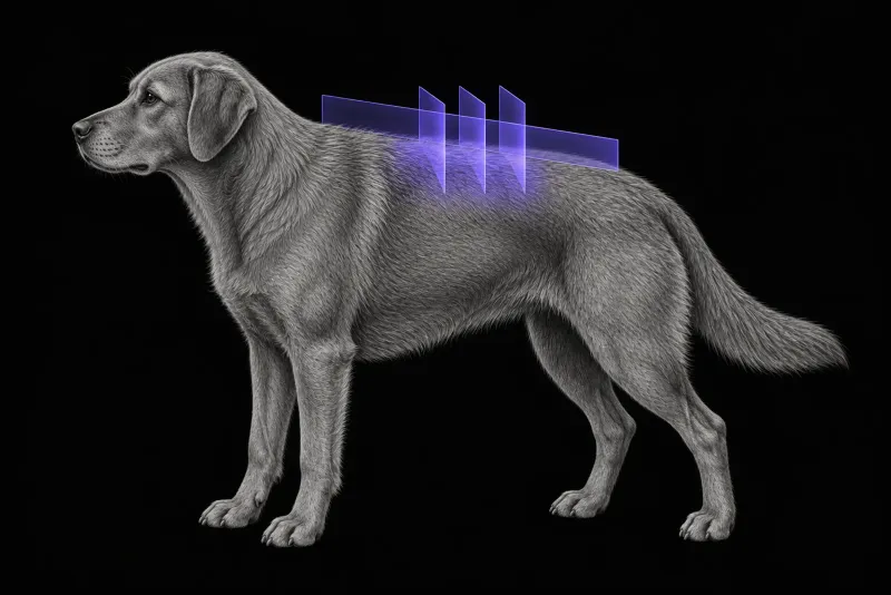
Why imaging decides the plan
Thoracolumbar intervertebral disc extrusion (TL-IVDE) is the most common cause of acute paraparesis and paraplegia in dogs, according to the ACVIM consensus statement on TL-IVDE (Olby et al., Journal of Veterinary Internal Medicine, 2022). The consensus recommends surgical management for non-ambulatory dogs that still have deep pain perception, and for paraplegic dogs that have lost it. Imaging confirms the diagnosis and also helps with surgical planning and prognosis.
Imaging also separates extrusion from protrusion and from other forms, as a 2020 review of imaging in disc disease (da Costa et al., Frontiers in Veterinary Science) describes:
- acute non-compressive nucleus pulposus extrusion (ANNPE)
- hydrated nucleus pulposus extrusion (HNPE)
- intradural or intramedullary extrusion
MRI, CT or myelography
The ACVIM consensus summarises the modalities as follows:
| Modality | What the consensus reports |
|---|---|
| MRI | Diagnostic sensitivity above 98.5%. Better than CT in dogs with peracute signs and for separating extrusion from protrusion. Shows residual compression after surgery. Adds prognostic information. Limits: longer acquisition, availability and cost. |
| CT | Rapid and lower cost. Sensitivity of 81% to 100% in chondrodystrophic dogs with mineralised discs. Less accurate in dogs older than 5 years and smaller than 7 kg. Does not show the severity of spinal cord injury, so its prognostic use is limited. |
| Myelography or CT-myelography | Sensitivity of 53% to 97%. Myelography is less accurate for lateralisation and carries a risk of seizures. |
The choice of modality can change the surgical plan. In 40 chondrodystrophic dogs that had both CT and MRI (Noyes et al., Veterinary Surgery, 2017), the same observer planned a similar hemilaminectomy on both studies in only 43.5% to 66.6% of dogs. The side was the most consistent element (87% to 92% agreement). Observers tended to plan larger defects on MRI.
Outcome data are less clear-cut. In a 2026 retrospective study of 560 non-ambulatory chondrodystrophic dogs (Low and Markovic, Veterinary Surgery), 42 of which were managed with CT-myelography, management guided by conditional CT-myelography was non-inferior to MRI for recovery of ambulation, with similar safety and lower cost.
For a young chondrodystrophic dog with a typical history, CT (with myelography when needed) is therefore a reasonable first step. MRI is usually the better choice when signs are peracute, when the dog is older than 5 years or lighter than 7 kg, or when prognosis is a main question, because CT is less accurate in those dogs and has limited prognostic use.
flowchart TD
  accTitle: CT or MRI for suspected thoracolumbar disc extrusion?
  accDescr: Based on the ACVIM consensus. MRI is usually the better choice for peracute signs, dogs older than 5 years or lighter than 7 kg, or when prognosis is a main question. Otherwise, in a young chondrodystrophic dog with a typical history, CT with myelography when needed is a reasonable first step.
  start["Suspected TL-IVDE"] --> q{{"Peracute, over 5 years, under 7 kg, or prognosis key?"}}
  q -- "Yes" --> mri["MRI"]
q -- "No, young chondrodystrophic dog" --> ct["CT, with myelography when needed"]Thoracolumbar spine MRI protocol at a glance
Under the ACVIM consensus, T2-weighted images are always included, and diagnostic accuracy suffers without transverse images. HASTE (half-Fourier single-shot turbo spin echo), T2* or STIR sequences may complement them. Contrast does not improve diagnostic accuracy or treatment planning in most cases.
What the radiologist looks at
- Extrusion type. In 51 dogs with HNPE compared with 105 dogs with TL-IVDE (Alcoverro et al., The Veterinary Journal, 2024), HNPE caused significantly less spinal cord compression. MRI-visible disc degeneration was present in every TL-IVDE case but in only 60% of HNPE cases.
- Intramedullary signal. The presence and extent of intramedullary T2 hyperintensity, T2 hypointensity, and loss of the cerebrospinal fluid (CSF) signal on HASTE or T2* sequences have been variably associated with a worse locomotor outcome and with progressive myelomalacia. Magnet strength, image plane and observer may affect how reliable T2 hyperintensity is as a prognostic sign.
- HASTE and deep pain. In 20 non-ambulatory dogs under 15 kg (Khan and Freeman, Veterinary Medicine and Science), the length of CSF signal loss on HASTE differed significantly between dogs that kept and dogs that lost deep pain perception. It did not differ significantly with neurological grade.
- Progressive myelomalacia. In 22 dogs with progressive myelomalacia and 38 paraplegic dogs without deep pain perception (Cordle et al., Veterinary Radiology & Ultrasound, 2023), the length of T2 hyperintensity and HASTE signal loss were associated with clinically evident myelomalacia. There was no significant difference when the analysis was limited to dogs not yet showing clinical signs of it.
- Other degenerate discs. In 92 dogs treated by hemilaminectomy alone (Longo et al., Journal of Veterinary Internal Medicine), a completely degenerate disc between T10 and L3, in addition to the operated site, was associated with a hazard ratio of 2.92 for recurrence of clinical signs.
What your report will include
Every Veheri case is read by a radiologist or neurologist, and when you order you can choose Diplomate-only reporting or the full specialist panel. Neurology reporting covers CT and MRI. You can chat directly with the specialist assigned to your case, and a simplified pet owner report helps you explain the findings to the owner.
When you take the report forward:
- Myelomalacia is not ruled out by a clear scan. Absence of T2 hyperintensity and HASTE signal loss supports absence of myelomalacia at the time of imaging, but does not reliably rule out imminent myelomalacia.
- Compression is not prognosis. In 17 dogs imaged before and immediately after surgery (Auffret et al., Veterinary Radiology & Ultrasound, 2024), residual extradural material was present in all postoperative studies. Neither its volume nor residual compression predicted the neurological outcome 2 to 5 weeks after surgery.
- Bladder outcome. Urinary outcomes were not associated with the severity or length of spinal cord compression on MRI, or with a focal versus extensive pattern.
- Mineralised discs cannot be specifically identified on MRI, and MRI findings support, but do not replace, the neurological grading of the dog.
- Talking to the owner: a completely degenerate disc elsewhere between T10 and L3 was associated with recurrence, which is worth mentioning when you discuss the longer term.
What to include when you request a thoracolumbar MRI report
- signalment: age, breed and sex
- weight, which with breed and age affects the choice between CT and MRI
- time of onset, and whether signs are peracute, progressive or static
- neurological grade, including deep pain perception, and the date of each examination
- any change since presentation, such as cranial movement of the cutaneous trunci reflex cut-off or loss of reflexes, which can point to progressive myelomalacia
- previous episodes, surgery or fenestration
- whether surgery is planned, so the report gives the site, side and extent of the extruded material
How Veheri can help
Veheri works with veterinary radiologists and neurologists and reports spinal MRI studies remotely for clinics. For the CT side of disc disease, see CT of the thoracolumbar spine in dogs, and for other presentations, see when veterinary MRI is the right move.
Sources
Each claim in this guide links to its source. The full citations:
- Olby NJ, Moore SA, Brisson B, et al. ACVIM consensus statement on diagnosis and management of acute canine thoracolumbar intervertebral disc extrusion. J Vet Intern Med. 2022;36(5):1570-1596. doi:10.1111/jvim.16480
- da Costa RC, De Decker S, Lewis MJ, et al. Diagnostic imaging in intervertebral disc disease. Front Vet Sci. 2020;7:588338. doi:10.3389/fvets.2020.588338
- Noyes JA, Thomovsky SA, Chen AV, et al. Magnetic resonance imaging versus computed tomography to plan hemilaminectomies in chondrodystrophic dogs with intervertebral disc extrusion. Vet Surg. 2017;46(7):1025-1031. doi:10.1111/vsu.12700
- Low D, Markovic A. Computed tomography versus magnetic resonance imaging for the perioperative management of acute thoracolumbar intervertebral disc extrusion in non-ambulatory chondrodystrophic dogs. Vet Surg. 2026;55(7):1198-1211. doi:10.1111/vsu.70135
- Alcoverro E, Schofield I, Spinillo S, et al. Thoracolumbar hydrated nucleus pulposus extrusion and intervertebral disc extrusion in dogs: Comparison of clinical presentation and magnetic resonance imaging findings. Vet J. 2024;306:106178. doi:10.1016/j.tvjl.2024.106178
- Khan S, Freeman P. HASTE MRI sequence findings correlate with loss of deep pain perception in dogs with thoracolumbar disc extrusion. Vet Med Sci. 9(2):604-608 (published online 2022). doi:10.1002/vms3.974
- Cordle KJ, Seiler GS, Barnes D, Olby NJ. MRI features can help to confirm a diagnosis of progressive myelomalacia, but may not be accurate in dogs lacking characteristic clinical signs at the time of imaging. Vet Radiol Ultrasound. 2023;64(2):283-293. doi:10.1111/vru.13178
- Longo S, Gomes SA, Briola C, et al. Association of magnetic resonance assessed disc degeneration and late clinical recurrence in dogs treated surgically for thoracolumbar intervertebral disc extrusions. J Vet Intern Med. 35(1):378-387 (published online 2020). doi:10.1111/jvim.15989
- Auffret V, Castel A, Juette T, et al. Residual volume of extruded disc material and residual spinal cord compression measured on postoperative MRI do not predict neurological outcomes in dogs following decompressive surgery for thoracolumbar intervertebral disc extrusion. Vet Radiol Ultrasound. 2024;65(2):76-86. doi:10.1111/vru.13325
Related reading
More imaging guides
All imaging guides
CT of the canine thoracolumbar spine for suspected disc extrusion
When non-contrast CT is enough for thoracolumbar disc extrusion in chondrodystrophic dogs, and which dogs need CT-myelography or MRI instead.
MRI of the feline thoracolumbar spine for disc disease
How MRI classifies disc extrusion, protrusion and non-compressive extrusion in cats, why degeneration is often incidental, and what outcome data show.
MRI of the canine brachial plexus for peripheral nerve sheath tumours
How MRI finds peripheral nerve sheath tumours of the canine brachial plexus, what the radiologist looks for, and what imaging can say about grade and extent.