
- Canine
- English cockerspaniel
- male, castrated
- 14 years old
One episode of being unable to urinate. A bleeding from the penis.
Patient
- Canine
- English cockerspaniel
- Male, castrated
- 14 years old
History
- One episode of being unable to urinate
- Bleeding from the penis
For further clarification, both radiographs and ultrasound images were taken.
Findings
Ultrasound
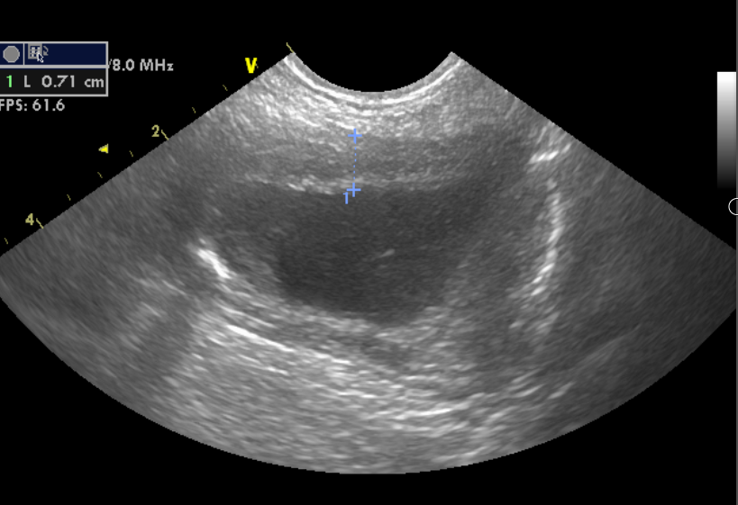
- Urinary bladder: Thicker wall (7 mm) with normal layering. A small hyperechoic line with a clean acoustic shadow at the level of the apex
- Kidneys were unremarkable (not included)
- Prostate: Symmetric, smooth margins, mildly enlarged (height 3 cm, width 4 cm), diffusely heteroechoic, multiple irregular hyperechoic lines with clean acoustic shadow. Mildly hyperechoic surrounding peritoneal fat. No free fluid was observed
- Medial iliac lymph node (MILn): Enlarged, hypoechoic, irregular nodular shape, hilus not visible
Radiographs
- Laterolateral and ventrodorsal views of the caudal abdomen and pelvis are available with very good quality (exposition and positioning). Images are centred for the spine and pelvis
- There is focal loss of serosal detail at the level of the prostate (hypogastrium). The prostate is poorly marginated with multiple small mineral opacities and dislocated cranially to the peritoneal cavity. The urinary bladder is slightly cranially dislocated and well marginated. The size and opacity are unremarkable
- No osteolysis or osteoproliferation of the pelvic bones, lumbar and sacral vertebrae is visible. Part of a metallic implant, likely from a previous surgery, is visible on the VD view lateral to the tibial plateau. Osteoproliferation of the lateral femoral condyle is visible
Conclusion/Diagnosis
- The most likely diagnosis is prostatic neoplasia
- The thickened urinary bladder wall indicates cystitis
- The hyperechoic sediment with a clean acoustic shadow corresponds to urocystoliths
- Medial iliac lymphadenopathy
- Differentials include metastatic and neoplastic, as well as reactive lymphadenopathy
Outcome
- The fine needle biopsy of the prostate was performed
- The cytologic conclusion was consistent with prostatic carcinoma
Discussion
- In castrated dogs, prostatic mineralisation observed on radiographs or ultrasound is a highly specific indicator of prostatic neoplasia, demonstrating 100% positive predictive value (PPV) and specificity, with a sensitivity of 84%. In contrast, in intact dogs without visible mineralisation, the likelihood of neoplasia is low (negative predictive value [NPV] of 96% and specificity of 77%). This emphasises the diagnostic significance of mineralisation in neutered dogs and highlights its limited predictive value in intact ones. This information is crucial for interpreting imaging findings in clinical decision-making
- The radiographs were taken after the ultrasound examination to check for potential bone metastasis, which accounts for the specific centring and collimation. It is essential to obtain both orthogonal views (laterolateral and ventrodorsal) to enhance the detection of bony changes. Thoracic radiographs should also be included in the assessment
- In this case, no regional bone lesions or distant metastases were identified. The osteoproliferation noted on the lateral femoral condyle is consistent with degenerative joint changes. Additionally, part of a metallic implant is visible lateral to the left knee, indicating a previous orthopaedic surgery that supports the diagnosis of degenerative joint changes
Further reading
Bradbury CA, Westropp JL, Pollard RE. Relationship between prostatomegaly, prostatic mineralisation, and cytologic diagnosis. Vet Radiol Ultrasound. 2009;50(2):167-171.