
- British Shorthair cat
- Female, intact
- 2 years old
- Presented with lethargy and anorexia
What is the underlying cause?
Case of the month September 2025 Patient and history
- British Shorthair cat
- Female intact
- 2 years old
- Presented with lethargy and anorexia
For further clarification, both radiographs and ultrasound images were taken.
Radiographic Description
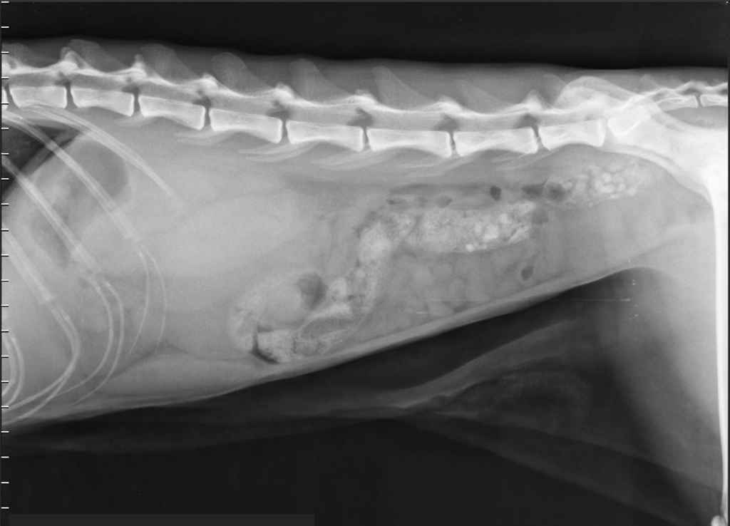
- Abdominal serosal detail is good. In the cranial and mid-retroperitoneal region, at the level of the kidneys, there are bilaterally poorly defined, ovoid, soft tissue-opaque masses (approximately 3–4 vertebral body lengths). Normal renal structures are not radiographically visible. The masses displace the cranial colon ventrally and caudally. The small intestinal loops are overall displaced ventrally and moderately filled with gas and fluid
- The stomach is moderately filled with gas and fluid and displaced slightly cranially. The colon contains gas and feces mixed with multiple mineral opacities
- The liver is not fully included in the image. The spleen and urinary bladder show no gross abnormalities, such as marked organomegaly, large masses, or mineralized calculi, within the limits of overlying intestinal loops. No skeletal abnormalities are detected
Ultrasound image
- The left kidney is markedly enlarged on ultrasound
- Normal corticomedullary differentiation is largely lost
- The renal surface is slightly irregular and shows an asymmetric, poorly defined hypoechoic rim
- The findings are suspicious for neoplastic infiltration, as can be seen with renal lymphoma
Diagnosis
- At least two large retroperitoneal masses are present
- The findings are most consistent with bilateral renomegaly due to neoplastic or inflammatory infiltration
- Another retroperitoneal pathology, such as hemangiosarcoma, is possible but considered less likely due to the bilaterally symmetric appearance
- A mesenteric mass is also considered unlikely
Outcome
- The masses were confirmed to originate from the kidneys
- Fine-needle aspiration revealed high-grade lymphocytic infiltration, consistent with renal lymphoma
- Chemotherapy resulted in temporary reduction of renal size and clinical improvement. However, the patient later developed a relapse and was euthanized
Discussion
- Renal lymphoma is the most common tumor of the kidneys in cats and one of the most frequent hematopoietic malignancies overall
- It can occur as a primary renal disease or as part of multicentric lymphoma
- Common clinical signs include anorexia, lethargy, weight loss, and changes in renal parameters (e.g., azotemia)
- Diagnosis is typically achieved via ultrasound and cytologic or histologic evaluation
- Prognosis depends on disease stage, presence of azotemia or anemia, and chosen therapy
- Chemotherapy can induce temporary improvement, but long-term survival is often limited