
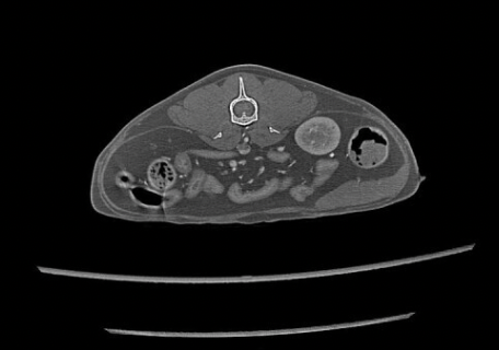
Cone Beam CT suits detailed dental, skull and small‑animal cases, with lower cost and dose. Conventional CT offers better soft tissue imaging and versatility, making it ideal for most veterinary practices.
Computed tomography (CT) has transformed veterinary imaging by improving diagnostic accuracy and expanding treatment options. As demand for advanced imaging grows, many veterinary practices are asking a key question:
Should we invest in a Cone Beam CT (CBCT) or a Conventional Multislice CT system?
Both systems provide three-dimensional imaging and high image quality, but they are fundamentally different in terms of applications, workflow, and diagnostic capabilities. Choosing the right CT scanner for your veterinary clinic depends on your case mix, specialisation, and long‑term goals.
Cone Beam CT vs. Conventional CT: What’s the Difference?
Veterinary practices typically choose between two main CT technologies:
- Conventional Multislice CT(also called fan-beam or helical CT)
- Cone Beam CT (CBCT), also known as Digital Volume Tomography (DVT)
Both are modern CT systems capable of excellent image quality, but they are optimised for different purposes.
- Conventional CT is designed for a wide range of indications and is considered the standard in general veterinary medicine and surgery.
- Cone Beam CT is highly specialised and excels in imaging very small, high‑contrast structures such as teeth, skull, and certain joints.
Most veterinary imaging experts agree that Conventional CT is the recommended choice for general practice with a broad caseload, while Cone Beam CT offers unique advantages for specific applications, particularly in dentistry and small exotic patients.
When Cone Beam CT Shines: Dentistry, Head Structures & Small Patients
Cone Beam CT provides major benefits when imaging small anatomical structures with high tissue density, especially in:
- Dentistry
- Oral and maxillofacial regions
- Certain joints
- Small mammals and exotic animals
Because of its technical design, CBCT delivers superimposition‑free, highly detailed 3D reconstructions, making it possible to visualise very fine anatomical details that may be missed or overlapped in conventional radiographs.
Key clinical advantages of Cone Beam CT in veterinary medicine:
- Head structures (dentistry, oral & maxillofacial cases)
- Small pets and exotic animals
- Joints and fine bony structures
Non‑Imaging Advantages of Cone Beam CT for Veterinary Practices
Beyond pure image quality in small structures, Cone Beam CT systems can offer several practical benefits for a clinic:
- Lower radiation dose compared with many Conventional CT protocols (Note: radiation dose is usually less critical in veterinary patients than in human medicine, but can still be a consideration.)
- Lower purchase price and operating costs
- Flexible reconstruction workflow
For practices focused primarily on dentistry, oral surgery, and small animal or exotic head imaging, a Cone Beam CT can be a cost‑effective solution that still delivers top‑tier diagnostic quality in its niche.
Beam Geometry and Contrast: Why It Matters for Soft Tissue Imaging
One of the most important technical differences between Cone Beam CT and Conventional CT relates to the X‑ray beam path and the resulting contrast resolution.
- Conventional CT uses a collimated, fan‑shaped beam
- Cone Beam CT uses a divergent, uncollimated cone‑shaped beam
In practical veterinary terms:
- In large dogs or larger body regions (thorax, abdomen), Cone Beam CT will usually provide inferior soft tissue contrast compared with a Conventional CT.
- In small patients or small regions (skull, limbs, small joints), the effect of scatter is less pronounced, and CBCT can perform very well—especially for high‑contrast structures like bone and teeth.
Clinical implication:
A Cone Beam CT is not ideal for routine thoracic, abdominal or soft tissue imaging in small or large animals.
Motion Artefacts: Impact on Image Quality and Workflow
Motion artefacts are a relevant concern in any CT modality, but they affect Cone Beam and Conventional CT differently.
- Cone Beam CT acquisition
- Conventional Multislice CT acquisition
For thoracic and abdominal exams, where breathing and organ motion are significant, Conventional CT is clearly less susceptible to motion artefactsand better suited for:
- Routine thoracic imaging
- Abdominal soft tissue evaluations
- Contrast‑enhanced examinations where timing is critical
Clear Limitations of Cone Beam CT in Veterinary Use
When evaluating CT systems for your veterinary practice, image quality in your most common indicationsshould be the top priority.
Cone Beam CT is a strong choice if:
- Your caseload is highly focused on:
- You rarely need to perform:
However, it is important to recognise the strong limitations of Cone Beam CT:
- Not suitable as a general‑purpose CT for body cavities
- Limited flexibility as your practice grows
If your practice frequently performs:
-
Thoracic CT (e.g. lung and mediastinal disease, metastasis checks)
-
Abdominal CT (e.g. oncology staging, complex abdominal cases)
-
Neurological, oncological or trauma cases requiring full‑body or multi‑region imaging …then you will need a multislice Conventional CT system to achieve:
-
High soft tissue contrast
-
Reliable thoracic and abdominal imaging
-
Reduced motion artefacts (especially with breath‑hold or controlled ventilation)
-
Broad diagnostic coverage for both small and large animals
In short: Conventional CT positions your practice much more broadly and supports a wide range of general medicine and surgical indications.
Teleradiology and Expert Interpretation: Another Practical Consideration
Another often‑overlooked factor is how easily your CT images can be interpreted by external specialists.
- Many veterinary radiologists and teleradiology providers primarily work with multislice Conventional CT.
- Cone Beam CT studies may be less common in their routine workflow and may not be accepted for all types of examinations.
- In many cases, Cone Beam CT images are only reported when used appropriately, such as for:
If your practice relies heavily on external teleradiology services, a Conventional CT will typically integrate more smoothly and provide more consistent diagnostic support across all body regions.
How to Decide: Cone Beam CT or Conventional CT for Your Veterinary Practice?
When choosing between Cone Beam and Conventional CT, consider the following questions:
- What are your main clinical indications?
- How important is soft tissue contrast in your day‑to‑day work?
- What level of future flexibility do you want?
- How will your images be interpreted?
Conclusion
Both Cone Beam CT and Conventional Multislice CT play important roles in modern veterinary medicine, but they are not interchangeable.
- Cone Beam CT
- Conventional Multislice CT
For most general medicine and surgery practices, a Conventional CT system is the more versatile and future‑proof option.For highly specialised dental or small‑exotic practices, a Cone Beam CT can be an excellent, focused solution with outstanding image quality in its niche.
Smarter Teleradiology. Faster Diagnosis. Better Results Join the Veheri Community!
Start today: Book your DEMO