
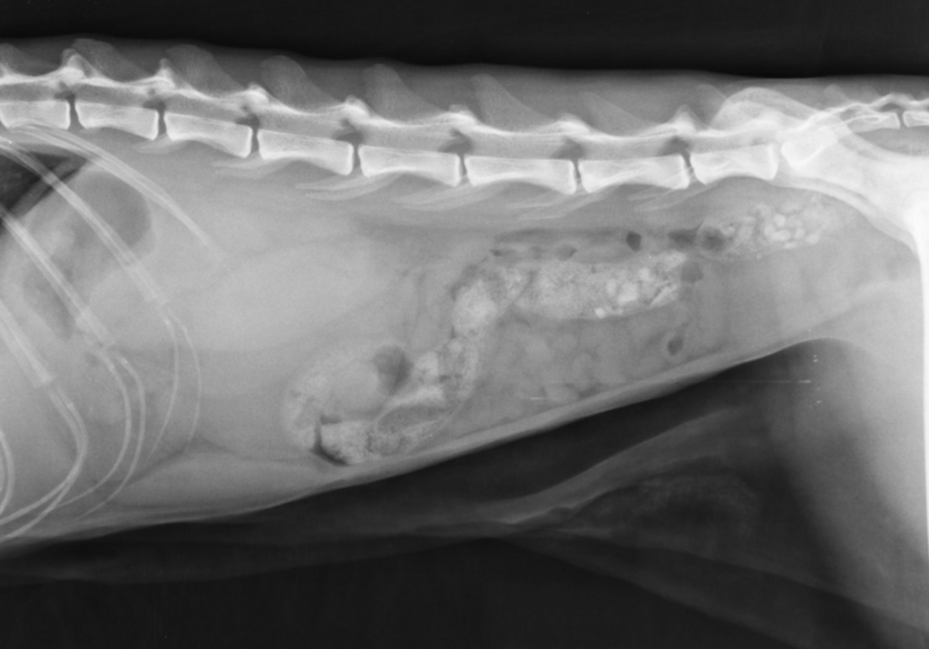
This guide delivers a modality-first framework, practical equipment specs, site‑planning and ALARA, workflow/PACS and teleradiology, and ROI modeling—focused only on CT, MRI, and X‑ray. With clear protocols and integration, you’ll improve diagnostic yield, safety, and throughput across global practice settings.
Veterinary diagnostic imaging solutions If you need a fast, practical answer: use X‑ray for first‑line skeletal and thoracic screening, CT for complex bone, sinus/nasal, chest, and trauma detail, and MRI for brain/spine and other soft‑tissue neuro‑musculoskeletal cases.
Modern veterinary teams need a clear, modality-first framework. This guide compares CT, MRI, and X‑ray for global practice settings, then gets tactical on equipment selection, room planning, safety (ALARA), workflow/PACS, and ROI. The goal is to help clinicians, managers, and procurement leads make confident, defensible choices that improve patient outcomes and throughput.
CT, MRI, and X‑ray at a glance
Choosing the right modality starts with clinical indication, patient stability, and logistics. X‑ray (radiography)is widely available, quick, and cost‑effective; CTadds cross‑sectional speed and outstanding bone and lung detail; MRIprovides unmatched soft‑tissue contrast, especially for neuro cases. Across worldwide markets, access and service coverage will influence feasibility as much as raw image quality.
- X‑ray (DR/CR):Two‑dimensional projection; ideal first‑line for fractures, thorax screening, and general orthopedics. Rapid, economical, and widely available. For client communication, X‑rays also serve as an accessible entry point before advanced imaging.
- CT: Cross‑sectional slices with superb bony detail and lung resolution; excellent for skull/nasal disease, ear, dental planning, thorax, trauma, and complex orthopedics; minutes to scan. Contrast studies and 3D reconstructions support surgery and radiation therapy planning.
- MRI:Superior soft‑tissue contrast; best for brain, spine, and soft‑tissue lesions (e.g., CNS, menisci/ligaments in select cases); runs longer and requires strict safety and anesthesia planning. Image quality relies on field strength, coils, and protocol design.
“CT and MRI play an essential role in cases where the benefit of the diagnostic yield outweighs the cost and necessity for general anesthesia.” — Matthew Wright, DVM, MS, DACVR via Today’s Veterinary Practice
Equipment selection: what matters for CT, MRI, and X‑ray
Clinically sound imaging begins with the right specifications. Modality decisions should match your case mix, throughput expectations, budget, and facility constraints. Spend as much time on serviceability and training as you do on headline specs—uptime and protocol quality are what the patient actually experiences.
- X‑ray:Prefer DR over CR for workflow speed and lower lifetime costs. Assess detector size (e.g., 14×17), tethered vs wireless, generator power (kW), and tables with floating tops for positioning. Consider grid use, exposure consistency (AEC), and compatibility with your PACS.
- CT:Slice count affects speed, resolution, and motion artifacts; 16‑slice systems are common entry points, while 32–64 slice improve throughput and image quality. Evaluate gantry aperture for large breeds, iterative reconstruction for dose/noise, and serviceability/parts availability in your region.
- MRI: Field strength and system design drive image quality and siting. High‑field (1.5T) systems offer superior neuro/orthopedic detail but demand rigorous siting, power, and quench infrastructure; dedicated veterinary/extremity systems can reduce footprint and cost. Confirm coil availability (neuro, MSK, body) and anesthesia‑safe workflows.
“In veterinary radiology we are currently in a digital radiography hotbed.” — Seth Wallack via dvm360
Site planning, safety, and ALARA
Every installation needs careful room preparation and written operating procedures. Shielding plans should be approved per local regulation; MRI rooms require controlled access, ferromagnetic screening, and quench/risk management; CT and X‑ray require radiation safety programs and personal dosimetry. Build culture and checklists around safety so that turnover or shift changes never compromise standards.
Radiation protection hinges on ALARA—protocol optimization, collimation, exposure charts, and staff training with appropriate PPE. Maintain up‑to‑date dose records and review repeats to find root causes. MRI safety demands zone controls, metal screening, and anesthesia‑compatible monitoring; enforce drills for quench and emergency scenarios.
For modality background and safety context, see Merck’s entries on CT in animals and MRI in animals.
Workflow, PACS, and teleradiology
Efficient imaging hinges on clean data pipelines. From acquisition to report, standardize DICOM tags, leverage PACS or VNA for archiving, and integrate with your PIMS for ordering and results. Version‑controlled protocols, consistent labeling, and structured reports reduce errors and accelerate care.
A reliable PACS accelerates comparisons, annotation, and multidisciplinary collaboration. Teleradiology partners extend subspecialty reads and off‑hours coverage; ensure SLA clarity on turnaround, urgent addendums, and discrepancy resolution. Build a QA/QC program with periodic protocol review, detector calibration, and image quality audits to sustain consistent performance.
Protocols, positioning, and quality
Consistent protocols reduce repeats, dose, and anesthesia time. For radiography, build species/region exposure charts and positioning guides with immobilization aids. For CT, create standard head, thorax, abdomen, and extremity protocols with contrast timing where applicable.
For MRI, define core sequences by region (e.g., brain: T2, FLAIR, T1 pre/post, GRE/SWI; spine: sagittal/axial T2/T1; MSK: PD/T2 fat‑sat) and sedate/anesthetize with motion in mind. Train teams on positioning landmarks, artifacts, and image acceptance criteria. Periodic peer review and protocol audits sustain image quality.
Budgeting, service, and ROI
Project total cost of ownership: purchase/lease, construction, shielding, anesthesia integration, service contracts, coils/detectors, downtime coverage, and training. Model utilization (cases/day), fee schedules, and payback horizon, remembering that consistent protocols and workflow often contribute more to ROI than raw hardware specs. In price‑sensitive regions, consider refurbished systems and OEM‑certified service where parts access and specialist engineers are limited.
Negotiate uptime SLAs, response times, and loaner coverage for critical components. Budget annually for QC, physics/engineering checks, and protocol refresh. Align pricing with case complexity to keep access equitable while protecting margins.
READ MORE at: Veterinary CT: What It Is, When Pets Need It, Safety, and Costs
Global market considerations
Supply chains, import rules, power quality, and service networks vary worldwide. Before procurement, validate parts logistics, field engineer coverage, and remote diagnostics in your country. For MRI, confirm helium supply and quench support; for CT and X‑ray, ensure generator serviceability and detector replacement timelines.
Align with regional radiation regulations and credentialing for staff exposure monitoring. Standardize training materials and SOPs across sites if you operate internationally, adapting to local regulatory language. When travel is limited, set up remote training and credentialing refreshers to preserve consistency.
Suggested visuals
Illustrations make modality selection and workflow tangible for teams and clients. Provide simple, labeled diagrams teams can revisit during onboarding and refreshers. Pair visuals with brief how‑to paragraphs so they stand on their own if printed.
- Modality decision matrix (indication vs modality): Provide a simple table mapping common indications to X‑ray/CT/MRI.
- Workflow diagram: Order → Acquisition → PACS → Teleradiology → Report → PIMS.
- ROI calculator snapshot: Inputs (cases/day, average fee, service cost) → Payback months.
Smarter Teleradiology. Faster Diagnosis. Better Results Join the Veheri Community! Start today: Book your DEMO