
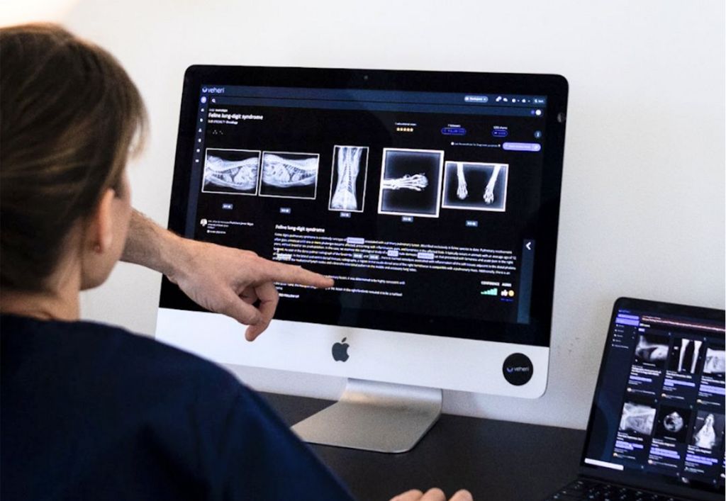
Navigating vet imaging diagnosis? Learn which diseases are best identified by CT, MRI, and X-rays, and how expert teleradiology can improve your clinical outcomes.
In the world of veterinary medicine, our patients can’t tell us where it hurts. As clinicians, we rely on vet imaging diagnosis to act as their voice. While a physical exam provides the “what,” diagnostic imaging provides the “where,” the “how much” and the “what next.”
Choosing between X-rays (Radiography), Computed Tomography (CT), and Magnetic Resonance Imaging (MRI)is one of the most critical decisions in a diagnostic workup. Each modality has its superpower in identifying specific diseases.
1. X-Rays: The Essential First Responder
Digital radiography remains the cornerstone of any initial vet imaging diagnosis. It is fast, widely available, and provides a “big picture” view of the patient.
- Orthopedic Injuries: Identifying bone fractures, luxations, and advanced osteoarthritis.
- Cardiopulmonary Disease: Detecting congestive heart failure (cardiomegaly), pneumonia, and pleural effusion.
- Gastrointestinal Obstructions: Spotting radiopaque foreign bodies or obstructive gas patterns.
- Uroliths: Visualizing bladder and kidney stones (though some are radiolucent and require further imaging).
2. CT Scans: The 3D Architect
When X-rays provide a flat, overlapping view, CT (Computed Tomography) breaks the body down into thin slices. It is the gold standard for complex anatomy and oncologic staging.
- Nasal and Ear Disease: Identifying chronic rhinitis, nasal tumors, and otitis media/interna.
- Oncology & Metastasis: CT is the most sensitive tool for detecting small pulmonary nodules (lung mets) and planning the surgical margin for complex masses.
- Vascular Anomalies: With contrast, CT is used to diagnose portosystemic shunts and other vascular malformations.
- Complex Fractures: Visualizing intra-articular fractures (like those in the elbow or tarsus) that are hidden on standard X-rays.
3. MRI: The Soft Tissue Specialist
While CT excels at bone and gas, MRI (Magnetic Resonance Imaging) is unrivaled for soft tissue contrast. If the problem is “hidden” inside the nervous system, MRI is the answer.
- Neurological Disorders: The primary tool for diagnosing Intervertebral Disc Disease (IVDD), brain tumors, encephalitis, and strokes.
- Soft Tissue Injuries: Identifying ligament tears (like the Cranial Cruciate Ligament) and tendonitis in high-performance or athletic animals.
- Spinal Cord Pathologies: Detecting syrinxes, cysts, or subtle nerve root compressions.
Which Modality Does Your Patient Need?
| Disease Suspected | Primary Modality | Why? |
|---|---|---|
| Bone Fracture | X-Ray / CT | Fast assessment (X-ray); 3D surgical planning (CT). |
| Seizures / Paralysis | MRI | Superior detail of brain and spinal cord tissue. |
| Nasal Discharge | CT | Excellent visualization of turbinate bone and sinuses. |
| Coughing / Dyspnea | X-Ray | Immediate screening for heart or lung changes. |
| Abdominal Mass | CT / Ultrasound | Determining organ involvement and vascular invasion. |
Optimize Your Diagnosis with Veheri
Acquiring the image is only half the battle; the true value of a vet imaging diagnosis lies in the interpretation. At Veheri, we connect veterinary clinics with world-class radiologists and neurologists to provide 24/7 teleradiology reports.
Start today: Book your DEMO Smarter Teleradiology. Faster Diagnosis. Better Results.
Imaging guides on this topic
Browse all imaging guides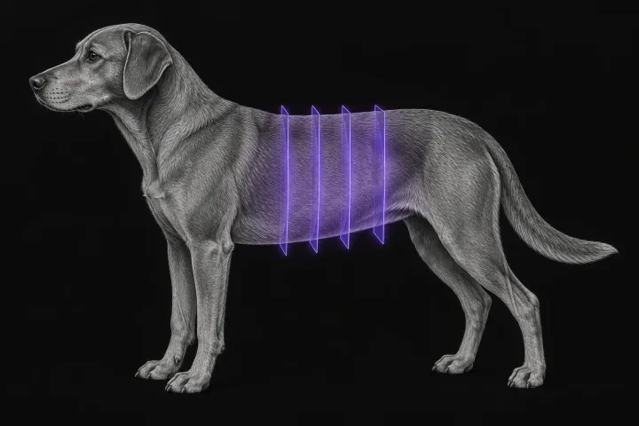
CT of the canine gastrointestinal tract in the acute abdomen
When abdominal CT adds to radiographs and ultrasound in vomiting dogs with suspected obstruction, foreign body or perforation, and what CT can and cannot show.
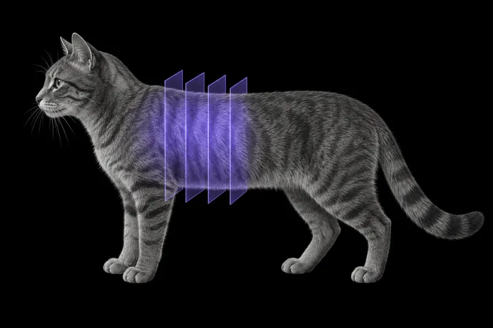
Thoracic CT in cats for lung tumours, airway and interstitial disease
What thoracic CT adds over radiographs in cats with lung tumours, bronchial or interstitial disease, how anaesthesia affects the lungs, and where CT stops.
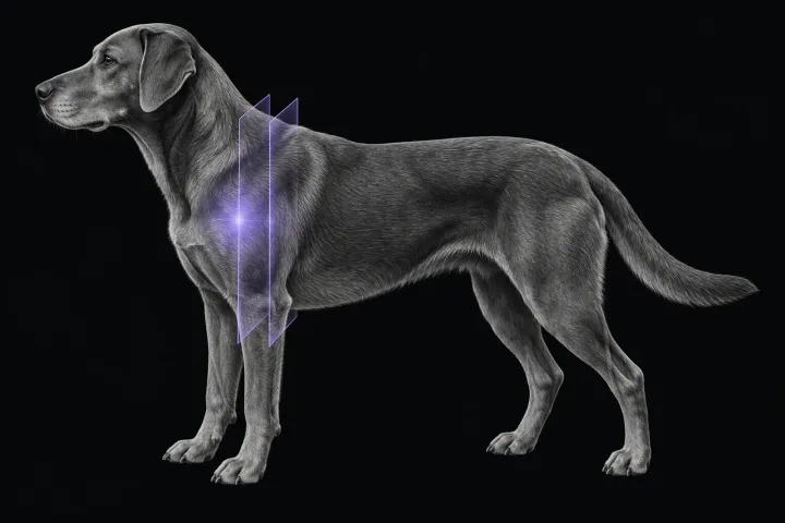
MRI of the canine shoulder for biceps and supraspinatus tendinopathy
What MRI shows in canine biceps and supraspinatus tendinopathy, how it compares with CT arthrography, and why a bright tendon alone is not disease.