
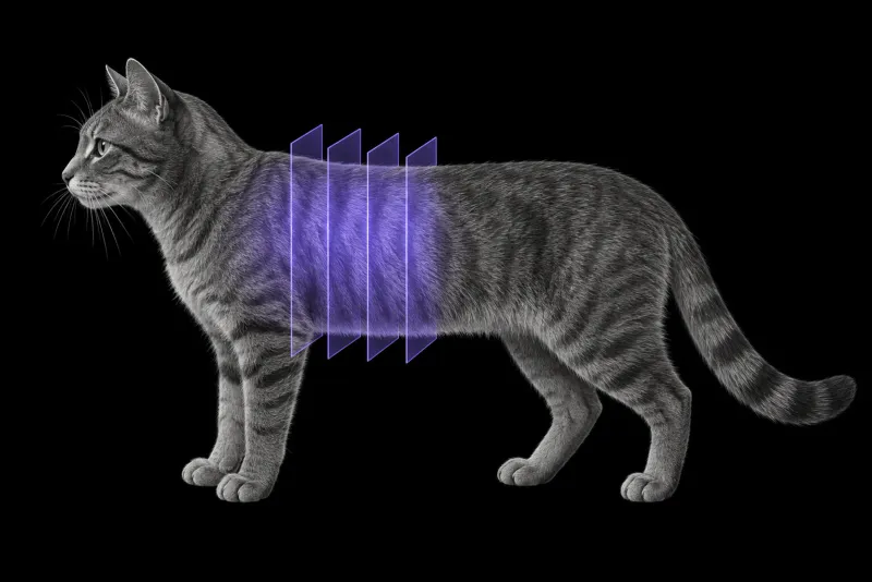
What thoracic CT adds over radiographs
In 54 cats with thoracic disease scanned awake in a positioning device (Oliveira et al., Veterinary Radiology & Ultrasound, 2011), 50 also had thoracic radiographs. The most common diagnoses were lung neoplasia, lower airway disease and cardiomyopathy (nine cats each).
- CT gave an additional correct diagnosis in 28% of cats (14 of 50) and additional information in 74% (37 of 50).
- Additional correct diagnoses were most common in lower airway disease.
- The extra CT findings were most often lung nodules, lung masses, bronchiectasis and mediastinal lymphadenopathy.
CT also finds smaller nodules. In cats with mammary carcinoma scanned awake (Klaengkaew et al., BMC Veterinary Research, 2021), awake CT found more cats with pulmonary nodules and smaller nodules than awake radiographs. The smallest nodule seen was 1.0 mm on CT and 2.5 mm on radiographs. CT also showed intrathoracic lymphadenopathy that radiographs did not.
CT agrees well with what the surgeon finds. In 64 dogs and cats that had thoracic surgery after CT (Brložnik et al., Frontiers in Veterinary Science, 2025), imaging and surgical findings agreed in 22 of 23 cats. Lung masses and pleural effusion showed complete agreement.
Bronchial and small-airway disease
- Bronchial wall thickening. In high-resolution CT of asthmatic and healthy cats (Won et al., Journal of Veterinary Medical Science, 2017), the ratios of bronchial wall thickness to bronchial diameter and to pulmonary artery size were higher in 4 asthmatic cats than in 16 healthy cats. The asthmatic group was small.
- Small airways. According to a consensus perspective on bronchiolar disorders in cats (Reinero et al., Journal of Veterinary Internal Medicine, 2019), thoracic radiography has low utility for bronchiolar disease. CT with paired inspiratory and expiratory scans can detect pathology centred on the small airways.
- Look upstream. In 549 cats with inflammatory upper airway disease or otitis media (Wetzels et al., Veterinary Radiology & Ultrasound, 2025), CT changes of lower airway disease were common. The odds ratio was 1.62 with rhinitis and 2.27 with rhinitis plus otitis media.
Feline thoracic CT protocol at a glance
How the cat is restrained changes the lungs. In healthy cats, general anaesthesia increased lung attenuation and was associated with atelectasis (odds ratio 15), especially in the cranial lobes, while awake CT in a positioning device was feasible, safe and clinically useful. A contrast series is worth including when a lung mass is known or suspected, small-airway questions call for paired inspiratory and expiratory scans, and when the head is scanned for nasal or ear disease, including the thorax is worth discussing.
What the radiologist looks at
- Atelectasis. Anaesthesia-related atelectasis affects lung interpretation, and awake scans have more motion artefact in the caudal thorax.
- A lung mass and everything around it. In 39 cats with primary pulmonary carcinoma examined post mortem (Santos et al., Veterinary Pathology, 2023), extrapulmonary metastases were present in 56.4%, most often in the regional lymph nodes (17 cats), skeletal muscles (9), kidneys (6) and parietal pleura (4). A larger primary tumour was associated with metastasis. The tumours appeared as a large nodule with smaller nodules, a solitary nodule, or multifocal to coalescent nodules. So the regional lymph nodes, every additional nodule and the pleura are described, not only the mass.
- Cavitary lesions. In 27 dogs and 15 cats with a gas-filled cavitary lung lesion (Parry et al., Journal of the American Veterinary Medical Association, 2023), six of 13 CT features were associated with the final diagnosis. Heterogeneous contrast enhancement, additional pulmonary nodules and a thicker lesion wall made malignancy more likely.
- Airway walls and small airways, using the bronchial ratios and paired scans above.
What your report will include
Every Veheri case is read by a radiologist or neurologist, and when you order you can choose Diplomate-only reporting or the full specialist panel. You can chat directly with the specialist assigned to your case, and a simplified pet owner report helps you explain the findings to the owner.
When you take the report forward:
- A fibrosis-like pattern is not a diagnosis. A 1-year-old cat with progressive signs and CT changes consistent with idiopathic pulmonary fibrosis, including traction bronchiectasis and architectural distortion, had no respiratory signs and resolution of the fibrosis-like CT patterns five months later (Stavri et al., BMC Veterinary Research, 2021). The authors suspected infection. They call for CT findings to be integrated with signalment, other clinical data and response to treatment before a diagnosis or prognosis of fibrotic disease is given in cats.
- Small and camouflaged lesions, such as low-contrast foreign bodies and ruptured bullae in atelectatic lung, were the ones most often missed or misinterpreted when CT was compared with surgery.
What to include when you request a thoracic CT report
- signalment: age, breed and sex
- respiratory signs, their duration, and any cough, tachypnoea or dyspnoea
- previous thoracic radiographs and echocardiography, if done
- known primary tumours elsewhere, for example mammary carcinoma
- concurrent nasal or ear disease
- whether the scan was awake, sedated or under general anaesthesia
How Veheri can help
Veheri’s radiologists report veterinary CT studies remotely for clinics, and offer expert support with CT protocols, positioning and acquisition. For related questions, see CT of the feline mediastinum and CT of the feline nasal cavity.
Sources
Each claim in this guide links to its source. The full citations:
- Oliveira CR, Mitchell MA, O'Brien RT. Thoracic computed tomography in feline patients without use of chemical restraint. Vet Radiol Ultrasound. 2011;52(4):368-76. doi:10.1111/j.1740-8261.2011.01814.x
- Brložnik M, Immler M, Prüllage ML, et al. Comparison of thoracic computed tomography and surgical reports in dogs and cats. Front Vet Sci. 2025;12:1566436. doi:10.3389/fvets.2025.1566436
- Klaengkaew A, Sutthigran S, Thammasiri N, et al. The evaluation of non-anesthetic computed tomography for detection of pulmonary parenchyma in feline mammary gland carcinoma: a preliminary study. BMC Vet Res. 2021;17(1):237. doi:10.1186/s12917-021-02950-6
- Santos IR, Raiter J, Lamego ÉC, et al. Feline pulmonary carcinoma: gross, histological, metastatic, and immunohistochemical aspects. Vet Pathol. 2023;60(1):8-20. doi:10.1177/03009858221122517
- Parry ME, Lumbrezer-Johnson S, Hostnik ET, et al. Cavitary pulmonary lesion wall thickness, presence of additional nodules, and intralesional contrast enhancement are associated with malignancy in dogs and cats. J Am Vet Med Assoc. 2023;261(10):1-7. doi:10.2460/javma.23.02.0076
- Won S, Yun S, Lee J, et al. High resolution computed tomographic evaluation of bronchial wall thickness in healthy and clinically asthmatic cats. J Vet Med Sci. 2017;79(3):567-571. doi:10.1292/jvms.16-0476
- Reinero CR, Masseau I, Grobman M, et al. Perspectives in veterinary medicine: description and classification of bronchiolar disorders in cats. J Vet Intern Med. 2019;33(3):1201-1221. doi:10.1111/jvim.15473
- Wetzels B, Ter Haar G, Hertog ED, Boroffka S. Computed tomographic evidence for united airway disease in cats: concurrent middle ear, upper and lower airway disease. Vet Radiol Ultrasound. 2025;66(3):e70032. doi:10.1111/vru.70032
- Stavri A, Masseau I, Reinero CR. Reversibility of clinical and computed tomographic lesions mimicking pulmonary fibrosis in a young cat. BMC Vet Res. 2021;17(1):380. doi:10.1186/s12917-021-03081-8
Related reading
More imaging guides
All imaging guides
Thoracic CT for pulmonary metastases in dogs
How thoracic CT compares with radiographs for pulmonary metastasis screening in dogs, which tumours most often spread to the lung, and how to read a nodule.
CT of the feline interscapular region for injection-site sarcoma
What contrast CT shows in feline injection-site sarcoma, how it changes surgical and radiotherapy planning, and where imaging cannot replace histology.
CT of the feline mediastinum for a cranial mediastinal mass
What CT adds for a cat with a cranial mediastinal mass, how far it separates thymoma, lymphoma and cysts, and why cytology or histology still decides.