
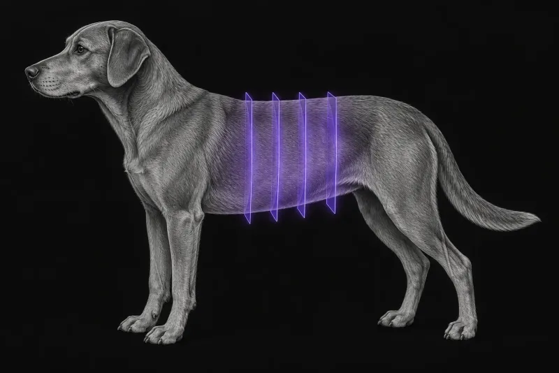
Radiographs and ultrasound come first
A vomiting dog with a suspected obstruction usually starts with radiographs, ultrasound or both. Both work, but each has gaps. In a comparison of radiography and ultrasound in 82 acutely vomiting dogs (Sharma et al., Veterinary Radiology & Ultrasound):
- a small-intestinal foreign body obstruction was confirmed in 27 (33%)
- radiography gave a definitive answer (obstructed or not) in 58 of 82 dogs (70%), and ultrasound in 80 of 82 (97%)
- about 30% of obstructed dogs (8 of 27) had no segmental small-intestinal dilatation on radiographs, and half of these had linear foreign bodies
In the same study, ultrasound had greater accuracy, fewer equivocal results and gave greater diagnostic confidence, and a jejunal diameter above 1.5 cm on ultrasound should prompt a careful search for a cause.
CT is usually worth discussing when radiographs and ultrasound remain equivocal, or when the surgeon wants the whole abdomen assessed before deciding.
What CT adds in suspected obstruction
- Accuracy. In 20 dogs reviewed by three experienced radiologists (Drost et al., Veterinary Radiology & Ultrasound, 2016), CT was more sensitive (95.8% vs 79.2%) and more specific (80.6% vs 69.4%) than radiographs for mechanical intestinal obstruction. The differences were not statistically significant. For recommending surgery, radiographs scored slightly higher than CT, again without a significant difference.
- Surgical confirmation. In 97 dogs and cats scanned for suspected obstruction (Miniter et al., PLOS ONE, 2019), CT found foreign material in 48 and mechanical obstruction in 41. In the 45 patients that went to laparotomy, the CT findings were confirmed in all.
- Surgical versus non-surgical disease. In 19 dogs with acute abdominal signs (Shanaman et al., Veterinary Radiology & Ultrasound, 2013), accuracy for separating surgical from non-surgical conditions was 100% for contrast-enhanced multidetector CT, 94% for ultrasound and 94% for radiography. Ultrasound underestimated the size and number of lesions compared with CT.
- Practical feasibility. In 18 dogs with acute abdominal signs scanned awake or lightly sedated (Shanaman et al., Veterinary Radiology & Ultrasound, 2012), dual-phase contrast CT was feasible. Median scan time was under 10 minutes. The two poor-quality scans were degraded by beam hardening from previously given barium and by motion artefact.
Ulcers, perforation and penetrating foreign bodies
In 82 dogs with confirmed gastrointestinal ulceration (Fitzgerald et al., Journal of Small Animal Practice, 2017), the sensitivities of each modality were:
| Radiography | Ultrasound | CT | |
|---|---|---|---|
| Non-perforated ulcer | 30% | 65% | 67% |
| Perforated ulcer | 79% | 86% | 93% |
Wooden skewers that leave the gut are a specific problem. In seven dogs with perforating wooden skewers (Costas and Cervera, Open Veterinary Journal, 2024), CT identified every skewer (median attenuation 79 HU). Most started from the stomach, then the jejunum.
Gastrointestinal CT protocol at a glance
The widely used approach for the acute abdomen is dual-phase contrast-enhanced CT. In a study of gastrointestinal wall conspicuity (Fitzgerald et al., Veterinary Radiology & Ultrasound, 2017), the wall was conspicuous in 56.7% of segments before contrast, 84.5% in the early postcontrast phase and 77.3% in the late phase, and the authors support dual-phase imaging. The scan can be done in awake or lightly sedated dogs, but barium given beforehand degrades the images.
What the radiologist looks at
- Obstruction and foreign material. Whether there is mechanical obstruction, where it is, and whether foreign material is visible, as in the 97-patient series.
- Signs of perforation. In perforated ulcers, the most frequent findings were peritoneal fluid (83%), a mural lesion (48%), peritoneal gas (31%) and a visible mucosal defect (29%).
- Peritoneal fat stranding. This was the most frequent finding around skewer perforations, while free fluid or gas was uncommon. Fat stranding was also seen on CT in dogs with gastric neoplasia with perforation, pancreatitis and small-intestinal foreign bodies.
- The pleural space. When a skewer crossed it, pleural effusion, pulmonary perforation and pneumothorax were present in most cases.
- A mass as the cause. In
41 dogs with gastrointestinal tumours (De Magistris et al., Veterinary Radiology & Ultrasound):
- epithelial tumours grew concentrically, spindle cell tumours eccentrically and round cell tumours in a mixed pattern
- only lymphomas were multifocal
- nine carcinomas and six spindle cell tumours caused partial stenosis with sub-obstruction
What your report will include
Every Veheri case is read by a radiologist or neurologist, and when you order you can choose Diplomate-only reporting or the full specialist panel. You can chat directly with the specialist assigned to your case, and a simplified pet owner report helps you explain the findings to the owner.
When you take the report forward:
- The decision to operate still rests on the whole picture. CT did not outperform radiography for this decision in one prospective study.
- Possible hypoperfused bowel or pancreas may need further imaging. Contrast-enhanced ultrasound detected perfusion deficits that CT missed, and the authors suggest focused contrast-enhanced ultrasound after CT may help identify hypoperfused lesions.
- A suspected tumour still needs histopathology. The CT growth patterns help rank the differentials; they do not replace it.
What to include when you request an abdominal CT report
- signalment: age, breed and sex
- duration of vomiting, abdominal pain and any known ingestion, including sticks, skewers or linear material
- findings of previous radiographs and ultrasound, with dates
- whether barium or other oral contrast has been given
- previous abdominal surgery
- the decision the CT has to support, for example surgery or continued monitoring
How Veheri can help
Veheri provides veterinary teleradiology and reports abdominal CT studies remotely for clinics. For the wider emergency picture, see the crucial role of emergency CT, and for routine abdominal scanning, our guide to abdominal CT protocols.
Sources
Each claim in this guide links to its source. The full citations:
- Sharma A, Thompson MS, Scrivani PV, et al. Comparison of radiography and ultrasonography for diagnosing small-intestinal mechanical obstruction in vomiting dogs. Vet Radiol Ultrasound. 52(3):248-55 (published online 2010). doi:10.1111/j.1740-8261.2010.01791.x
- Drost WT, Green EM, Zekas LJ, et al. Comparison of computed tomography and abdominal radiography for detection of canine mechanical intestinal obstruction. Vet Radiol Ultrasound. 2016;57(4):366-75. doi:10.1111/vru.12353
- Miniter BM, Gonçalves Arruda A, Zuckerman J, et al. Use of computed tomography (CT) for the diagnosis of mechanical gastrointestinal obstruction in canines and felines. PLoS One. 2019;14(8):e0219748. doi:10.1371/journal.pone.0219748
- Shanaman MM, Schwarz T, Gal A, O'Brien RT. Comparison between survey radiography, B-mode ultrasonography, contrast-enhanced ultrasonography and contrast-enhanced multi-detector computed tomography findings in dogs with acute abdominal signs. Vet Radiol Ultrasound. 2013;54(6):591-604. doi:10.1111/vru.12079
- Shanaman MM, Hartman SK, O'Brien RT. Feasibility for using dual-phase contrast-enhanced multi-detector helical computed tomography to evaluate awake and sedated dogs with acute abdominal signs. Vet Radiol Ultrasound. 2012;53(6):605-12. doi:10.1111/j.1740-8261.2012.01973.x
- Fitzgerald E, Barfield D, Lee KC, Lamb CR. Clinical findings and results of diagnostic imaging in 82 dogs with gastrointestinal ulceration. J Small Anim Pract. 2017;58(4):211-218. doi:10.1111/jsap.12631
- Costas A, Cervera V. CT findings in seven dogs with perforating wooden skewer foreign bodies from the gastrointestinal tract. Open Vet J. 2024;14(4):1002-1011. doi:10.5455/OVJ.2024.v14.i4.7
- Fitzgerald E, Lam R, Drees R. Improving conspicuity of the canine gastrointestinal wall using dual phase contrast-enhanced computed tomography: a retrospective cross-sectional study. Vet Radiol Ultrasound. 2017;58(2):151-162. doi:10.1111/vru.12467
- De Magistris AV, Rossi F, Valenti P, et al. CT features of gastrointestinal spindle cell, epithelial, and round cell tumors in 41 dogs. Vet Radiol Ultrasound. 64(2):271-282 (published online 2022). doi:10.1111/vru.13188
Related reading
More imaging guides
All imaging guides
CT of adrenal masses in dogs
What contrast CT shows in a dog with an adrenal mass, from caval invasion and adhesions to tumour type, and where its accuracy runs out before adrenalectomy.

Brain CT in dogs when MRI is not available
What brain CT can and cannot detect in dogs when MRI is not available, for masses, stroke, inflammation and head trauma, and when MRI or CSF is still needed.
CT of the canine elbow for medial coronoid disease
When CT is worth it for medial coronoid disease in dogs, what it finds that radiographs miss, and where arthroscopy still gives the better answer.