
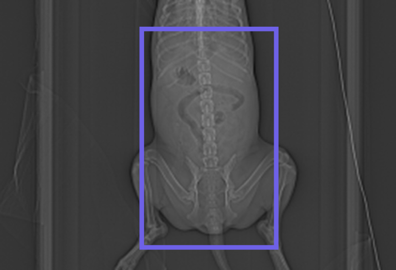
In modern veterinary diagnostics, a standardized Abdominal Routine CT is a cornerstone for accurate clinical assessment. This protocol is primarily indicated for screening primary neoplasia or metastasis, identifying foreign bodies, evaluating trauma, and detailed surgical planning.
In the rapidly evolving field of veterinary diagnostics, Computed Tomography (CT) has shifted from a luxury to a necessity for high-standard clinical practice. For practitioners looking to elevate their diagnostic accuracy, understanding the nuances of an Abdominal Routine Protocol is essential for identifying everything from subtle neoplastic changes to acute traumatic injuries.
The following guide, based on the Veheri standards, outlines the optimal workflow for consistent, high-quality abdominal imaging.
1. Clinical Indications: When to Scan?
An abdominal CT is a powerful tool, but its utility depends on the clinical context. Standard protocols are primarily recommended for:
- Oncology: Primary neoplasia identification and meticulous metastatic screening.
- Emergency & Trauma: Evaluating internal injuries and surgical planning following acute trauma.
- Gastrointestinal Issues: Precise localization of foreign bodies.
- Pre-surgical Mapping: Providing surgeons with a 3D understanding of vascular and organ involvement.
2. Patient Preparation: Setting the Stage for Success
Quality imaging begins long before the X-ray tube starts spinning. Proper positioning and motion control are the “make or break” factors in veterinary CT.
Positioning Strategy
The patient must be placed in dorsal recumbency. To prevent “superposition” (where limbs obscure the internal organs), the pelvic limbs should be caudally extended and abducted. This moves the dense bone structure out of the field of view, allowing for a cleaner scan of the caudal abdomen and pelvic inlet.
Achieving “Zero” Motion
Motion artifact is the enemy of diagnostic clarity. To combat this, the protocol suggests hyperventilating the patientimmediately prior to the scan to induce a temporary state of apnoea. This ensures the diaphragm remains still, preventing the “blurring” of cranial abdominal organs like the liver and spleen.
3. Technical Parameters & Tailored Dosing
Not all patients are created equal, and your CT settings shouldn’t be either. The protocol scales based on the patient’s weight to ensure adequate penetration without unnecessary radiation.
| Breed/Size Category | Weight | kV | mAs | Slice Thickness |
|---|---|---|---|---|
| Toy/Small | < 15 kg | 120 kV | 160 mAs | 1.5 mm |
| Medium | 16-30 kg | 120 kV | 160 mAs | 2.5 mm |
| Large/Giant | > 30 kg | 120 kV | 200 mAs | 3 mm |
All protocols utilize a 1-second rotation time and a pitch ranging from 1.4 to 2.0 depending on the size of the animal.
4. The Role of Contrast Media
While a pre-contrast scan (Phase 1) is necessary for a baseline, the post-contrast scan (Phase 2) is vital when inflammatory disease or neoplasia is suspected.
- Dose: $2ml/kg$ of Iohexol ($300/350
mgl/ml$). - Access: It is critical to maintain I/V access throughout the entire procedure.
- Timing: The “sweet spot” for routine abdominal enhancement is exactly 1 minute post-injection. At this stage, you should verify contrast visibility in the aorta to confirm a successful vascular phase.
5. Reconstruction and Submission
The final step is translating raw data into readable images. The Veheri protocol mandates specific reconstruction algorithms for the transverse plane:
-
Pre-Contrast: Submit both Osteo/Sinus (Sharp) for bone detail and Soft Tissue/Mediastinum (Soft) for organ parenchyma.
-
Post-Contrast: Generally requires only the Soft Tissue/Mediastinum reconstruction.
Disclaimer: While these parameters serve as a professional suggestion, every CT system has unique requirements. Always consult with your application specialist to optimize these settings for your specific hardware.
[Download the Complete Protocol Now] Start today: Book your DEMO Smarter Teleradiology. Faster Diagnosis. Better Results.
Imaging guides on this topic
Browse all imaging guides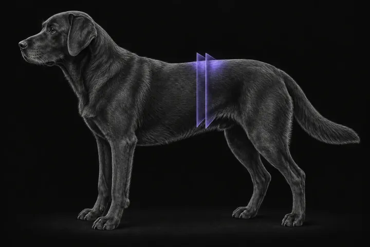
CT of adrenal masses in dogs
What contrast CT shows in a dog with an adrenal mass, from caval invasion and adhesions to tumour type, and where its accuracy runs out before adrenalectomy.
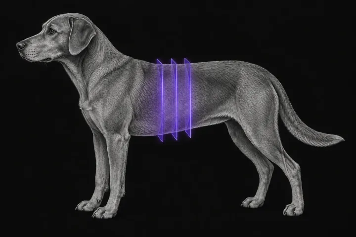
Multiphase CT of the canine liver for focal masses
How multiphase CT characterises a focal liver mass in dogs, how reliable its benign versus malignant signs are, and what it adds to surgical planning.
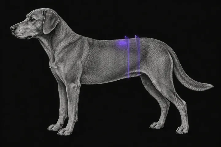
CT of the sublumbar lymph nodes for anal sac adenocarcinoma staging
How CT compares with abdominal ultrasound for staging the iliosacral lymph nodes in dogs with anal sac adenocarcinoma, and what the report should state.