
Why the iliosacral lymph nodes decide the plan
Anal sac adenocarcinoma spreads early to the regional lymph nodes, and a small primary tumour does not rule this out:
- In 116 dogs with abdominal staging and cytological or histological confirmation (Jones and Wustefeld-Janssens, Veterinary and Comparative Oncology, 2023), 53 (46%) had lymph node metastasis at presentation. The rate was 20% (9 of 46) for primary tumours under 2 cm and 63% (44 of 70) for tumours of 2 cm or more.
- In 70 dogs staged with CT of the thorax, abdomen and pelvis (Sutton et al., Journal of Small Animal Practice, 2022), iliosacral lymphadenomegaly was present in 71% and pulmonary metastases in 11%. Anal sac mass size was not associated with lymph node enlargement. No dog had pulmonary metastases without locoregional lymphadenomegaly.
- Calcium is not a substitute for imaging. In 58 dogs (Toth et al., Veterinary Medicine and Science, 2024), hypercalcaemia was associated with confirmed or suspected iliosacral metastasis, but 11 of 24 dogs (46%) with nodal disease were normocalcaemic.
The number of metastatic nodes also matters. In 57 dogs treated with sacculectomy and lymphadenectomy (Tanis et al., Veterinary and Comparative Oncology, 2022), more than four metastatic nodes was associated with shorter progression-free interval and disease-specific survival. The size of the largest node was not associated with outcome, and its measurement on imaging was not consistently reproducible.
The anatomy that makes staging difficult
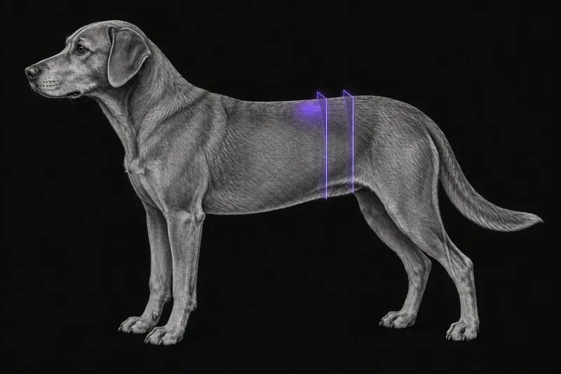
The iliosacral lymph centre consists of the medial iliac, internal iliac (formerly hypogastric) and sacral lymph nodes. The sacral nodes lie inside the pelvis, ventral to the sacrum and dorsal to the pubis. According to a comparison of CT and ultrasound in 20 dogs (Palladino et al., Journal of Veterinary Internal Medicine, 2016), this makes them hard to image with ultrasound, and they can be missed at exploratory surgery. In a lymphoscintigraphy study in eight healthy dogs (Linden et al., Veterinary Radiology & Ultrasound, 2019), a sacral node was the sentinel node in three (37.5%), and the injection technique changed which node was identified.
Ultrasound, CT and MRI compared
| Study | What it found |
|---|---|
| CT vs ultrasound, 12 dogs, prospective (Pollard et al., Veterinary and Comparative Oncology, 2017) | CT identified more iliosacral nodes (61 vs 30), more medial iliac nodes (33 vs 19) and more sacral nodes (15 vs 0). Ultrasound identified slightly more abnormal nodes (7 vs 5). |
| CT vs ultrasound, 20 dogs, retrospective (Palladino et al.) | CT found lymphadenomegaly in 13 dogs. Ultrasound found at least one enlarged node in all 13, but found every enlarged node in only 4 (30.8%). It detected 7 of 8 enlarged medial iliac, 4 of 11 internal iliac and 4 of 9 sacral nodes. |
| MRI vs ultrasound, 6 dogs with confirmed nodal metastasis (Anderson et al., Veterinary and Comparative Oncology, 2015) | MRI showed lymphadenopathy in all 6 dogs; ultrasound in 2. Lymphadenopathy was mainly sacral. |
The authors of the larger CT study concluded that abdominal ultrasound is an effective screening test for iliosacral lymphadenomegaly, but CT should be considered when an additional metastatic site would change the treatment plan. They note that identifying which specific nodes are enlarged may matter for surgical and radiotherapy planning.
Sublumbar CT protocol at a glance
Staging CT that covers the thorax, abdomen and pelvis, as in the 70-dog staging study, includes the lungs and every iliosacral node group, including the intrapelvic sacral nodes. Sacral nodes tend to be the smallest in this group, and thinner slices may improve measurement accuracy.
What the radiologist looks at
- Each node group on each side. Medial iliac, internal iliac and sacral nodes, and which of them are enlarged.
- Size against a reference. There are no pathognomonic imaging signs of nodal metastasis, so size is the usual criterion. The 20-dog study normalised node diameter to the aortic diameter at L5–L6. Using 30 presumed normal dogs as a reference, it classed a node as enlarged at a node-to-aorta ratio of at least 1.3 for medial iliac nodes and at least 1.0 for internal iliac and sacral nodes.
- The number of abnormal nodes, because more than four metastatic nodes was associated with shorter survival.
- The lungs, when the thorax is included.
What your report will include
Every Veheri case is read by a radiologist or neurologist, and when you order you can choose Diplomate-only reporting or the full specialist panel. You can chat directly with the specialist assigned to your case, and a simplified pet owner report helps you explain the findings to the owner.
When you take the report forward:
- Enlarged does not prove metastatic. In the 20-dog study only 5 dogs had nodes sampled, and all 5 were metastatic. The authors recommend sampling mildly to moderately enlarged nodes by ultrasound-guided aspiration or core biopsy.
- Surgical and radiotherapy planning may depend on which specific nodes are enlarged, not only on whether any are.
- Normal calcium does not rule out nodal disease: 11 of 24 dogs (46%) with nodal disease were normocalcaemic.
What to include when you request a CT report
- signalment: age, breed and sex
- side and measured size of the anal sac mass, and rectal examination findings
- cytology or histology of the primary tumour and any nodes sampled
- calcium results
- previous abdominal ultrasound findings, so the two studies can be compared
- the planned treatment: sacculectomy, lymphadenectomy, radiotherapy or medical therapy
How Veheri can help
Veheri’s radiologists report veterinary CT studies remotely for clinics, and offer expert support with CT protocols, positioning and acquisition. For lung staging, see thoracic CT for pulmonary metastases in dogs, and for the wider picture, our oncology imaging guide.
Sources
Each claim in this guide links to its source. The full citations:
- Jones AE, Wustefeld-Janssens BG. A relatively high proportion of dogs with small apocrine gland anal sac adenocarcinoma (AGASACA) primary tumours present with locoregional lymph node metastasis. Vet Comp Oncol. 2023;21(2):327-331. doi:10.1111/vco.12890
- Sutton DR, Hernon T, Hezzell MJ, et al. Computed tomographic staging of dogs with anal sac adenocarcinoma. J Small Anim Pract. 2022;63(1):27-33. doi:10.1111/jsap.13426
- Toth D, Upchurch D, Hallman RM. Association between total hypercalcaemia and iliosacral lymph node metastasis in dogs diagnosed with anal sac adenocarcinoma using abdominal ultrasonography. Vet Med Sci. 2024;10(1):e1324. doi:10.1002/vms3.1324
- Tanis JB, Simlett-Moss AB, Ossowksa M, et al. Canine anal sac gland carcinoma with regional lymph node metastases treated with sacculectomy and lymphadenectomy: outcome and possible prognostic factors. Vet Comp Oncol. 2022;20(1):276-292. doi:10.1111/vco.12774
- Palladino S, Keyerleber MA, King RG, et al. Utility of computed tomography versus abdominal ultrasound examination to identify iliosacral lymphadenomegaly in dogs with apocrine gland adenocarcinoma of the anal sac. J Vet Intern Med. 2016;30(6):1858-1863. doi:10.1111/jvim.14601
- Linden DS, Cole R, Tillson DM, et al. Sentinel lymph node mapping of the canine anal sac using lymphoscintigraphy: a pilot study. Vet Radiol Ultrasound. 2019;60(3):346-350. doi:10.1111/vru.12707
- Pollard RE, Fuller MC, Steffey MA. Ultrasound and computed tomography of the iliosacral lymphatic centre in dogs with anal sac gland carcinoma. Vet Comp Oncol. 2017;15(2):299-306. doi:10.1111/vco.12160
- Anderson CL, MacKay CS, Roberts GD, et al. Comparison of abdominal ultrasound and magnetic resonance imaging for detection of abdominal lymphadenopathy in dogs with metastatic apocrine gland adenocarcinoma of the anal sac. Vet Comp Oncol. 2015;13(2):98-105. doi:10.1111/vco.12022
Related reading
More imaging guides
All imaging guides
CT of adrenal masses in dogs
What contrast CT shows in a dog with an adrenal mass, from caval invasion and adhesions to tumour type, and where its accuracy runs out before adrenalectomy.

Brain CT in dogs when MRI is not available
What brain CT can and cannot detect in dogs when MRI is not available, for masses, stroke, inflammation and head trauma, and when MRI or CSF is still needed.
CT of the canine elbow for medial coronoid disease
When CT is worth it for medial coronoid disease in dogs, what it finds that radiographs miss, and where arthroscopy still gives the better answer.