
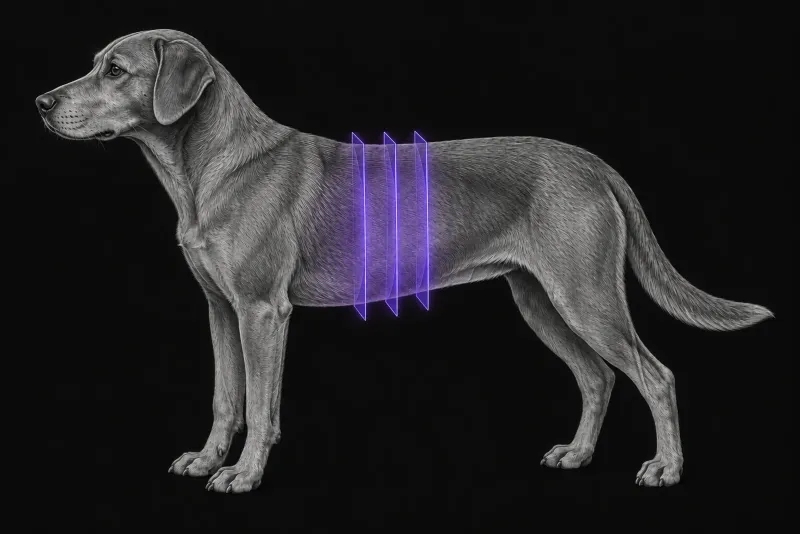
The questions a liver mass raises
When ultrasound finds a focal liver mass, the next decisions depend on three questions:
- is it likely to be malignant?
- where exactly is it?
- can it be removed?
Multiphase CT can contribute to all three, with limits that the report should state plainly.
Benign or malignant: what multiphase CT shows
Several studies have compared CT patterns with histopathology. The most consistent findings come from a triple-phase CT study of dogs with hepatic masses (Kutara et al., Veterinary Radiology & Ultrasound) and a study of CT characteristics of primary hepatic masses (Fukushima et al., Veterinary Radiology & Ultrasound, 2012):
| Lesion | CT features reported |
|---|---|
| Hepatocellular carcinoma | Heterogeneous enhancement with mixed hyper-, iso- and hypoenhancement in the arterial and portal phases (40 of 47, 85.1%) (Kutara et al.). Central and marginal arterial enhancement, cyst-like areas and a capsule, with hypoattenuation in the portal and equilibrium phases (Fukushima et al.). |
| Nodular hyperplasia | Homogeneous hyper- or isoenhancement in the portal and delayed phases (13 of 14, 92.9%) (Kutara et al.). Less often encapsulated and smaller than carcinoma or adenoma (Fukushima et al.). |
| Hepatocellular adenoma | Diffuse arterial enhancement in 57%, a pattern also seen in nodular hyperplasia but never in carcinoma (Fukushima et al.). |
| Metastatic tumour | Homogeneous hypoenhancement in the arterial and portal phases (8 of 9, 88.9%) (Kutara et al.). |
Quantitative cut-offs. In 94 dogs with histologically diagnosed liver lesions (Einwaller and Wennemuth, Veterinary Radiology & Ultrasound, 2025), hepatocellular carcinomas were larger and enhanced heterogeneously. In the portal venous phase, benign lesions were hyperattenuating to the liver and carcinomas hypoattenuating. Three features were associated with carcinoma, with an accuracy of 89% and a positive predictive value of 91%:
- a maximum transverse diameter above 9.8 cm
- heterogeneous enhancement in all three phases
- a portal venous attenuation below 136 HU
In another study of 46 masses in 44 dogs (Griebie et al., American Journal of Veterinary Research, 2017), a delayed-phase absolute enhancement below 37 HU suggested malignancy and correctly classified 42 of 46 masses (91.3%). Triphasic CT was more accurate than ultrasound, which reached 80.4% when cavitated masses were called malignant.
Pooled evidence. A meta-analysis (Burti et al., The Veterinary Journal, 2021) identified these features as informative:
- well-defined margins
- a capsule
- abnormal lymph nodes
- heterogeneity in the arterial, portal and delayed phases
Pooled sensitivity ranged from 0.630 to 0.786 and pooled specificity from 0.643 to 0.816. Larger size and lower attenuation values were more associated with malignancy.
A necessary caution: the patterns overlap
Not every study found clear separation. In 24 hepatic masses examined with dual-phase CT (Jones et al., Veterinary Radiology & Ultrasound, 2016), there was substantial overlap between malignant and non-malignant masses. Most hepatic masses showed marked generalised early enhancement that persisted into the delayed phase, whatever the histology. The authors concluded that dual-phase CT gave limited specific diagnostic information.
Read together, these studies suggest that a combination of several features carries more weight than any single sign. A CT pattern raises or lowers the likelihood of malignancy; it is not a histological diagnosis.
Where is the mass, and can it be removed?
- Location. In 71 dogs with a solitary mass confirmed at surgery or necropsy (Cheney et al., Journal of Small Animal Practice, 2019), location was correctly predicted in 42 of 57 dogs (74%) by ultrasound and in 37 of 44 (84%) by CT. Both had high specificity, and sensitivity was highest for left division masses. The authors consider either modality appropriate for presurgical planning.
- Resectability. In a prospective study of 20 dogs with 21 isolated liver masses (Smola et al., Journal of the American Veterinary Medical Association, 2023), CT angiography was reviewed by a surgeon and by a blinded radiologist. Seventeen masses were grossly resectable, and two more that seemed resectable were incompletely excised on histopathology. Both reviewers predicted gross resectability and complete excision accurately. The radiologist localised lesions more accurately; the surgeon better predicted surgical difficulty.
Liver CT protocol at a glance
Most of the studies above used contrast-enhanced multiphase CT. Triple-phase imaging covers the arterial, portal venous and delayed phases, as in the triple-phase study of hepatic masses. The dual-phase study used two phases instead, and CT angiography was used to judge resection in the resectability study.
What the radiologist looks at
- Enhancement pattern in each phase. Homogeneous or heterogeneous, and hyper-, iso- or hypoattenuating to the liver, weighed together rather than as single signs, because the patterns overlap.
- Size, margins and attenuation. Larger size, lower attenuation values, margins and a capsule all feature in the pooled evidence.
- Location. The affected lobe or division, since sensitivity for localisation was highest for left division masses.
- Vessels and lobes. The relationship to the hepatic veins, portal vein and caudal vena cava, and the number of lobes involved, because major vascular involvement, multilobar involvement and right-sided location reduced resectability.
- Regional lymph nodes. Abnormal lymph nodes were among the informative features in the meta-analysis.
What your report will include
Every Veheri case is read by a radiologist or neurologist, and when you order you can choose Diplomate-only reporting or the full specialist panel. You can chat directly with the specialist assigned to your case, and a simplified pet owner report helps you explain the findings to the owner.
When you take the report forward:
- A suggested benign or malignant pattern shifts the probability; it does not replace histopathology, because benign and malignant masses can enhance alike.
- A mass that looks resectable may still be incompletely excised: two masses that seemed resectable were incompletely excised on histopathology.
- Metastatic disease stays on the differential list when there is neoplasia elsewhere, since metastases showed their own enhancement pattern.
- Talking to the owner: CT can make malignancy more or less likely and help plan surgery, but the final diagnosis comes from histopathology.
What to include when you request a liver CT report
- signalment: age, breed and sex
- ultrasound findings, including size, location and any cavitation
- whether surgery is being considered, so resectability can be addressed
- any history of neoplasia elsewhere, since metastases are part of the differential
- relevant blood test results and any previous sampling of the mass
- whether thoracic staging should be included in the same study
How Veheri can help
Veheri’s radiologists report veterinary CT studies remotely for clinics, and can support your team with CT scanning protocols. For related reading, see CT of splenic masses in dogs and oncology imaging for staging and planning.
Sources
Each claim in this guide links to its source. The full citations:
- Kutara K, Seki M, Ishikawa C, et al. Triple-phase helical computed tomography in dogs with hepatic masses. Vet Radiol Ultrasound. 55(1):7-15 (published online 2013). doi:10.1111/vru.12099
- Fukushima K, Kanemoto H, Ohno K, et al. CT characteristics of primary hepatic mass lesions in dogs. Vet Radiol Ultrasound. 2012;53(3):252-7. doi:10.1111/j.1740-8261.2011.01917.x
- Einwaller J, Wennemuth J. Diagnostic utility of triple-phase CT: differentiating benign liver lesions from hepatocellular carcinoma in dogs. Vet Radiol Ultrasound. 2025;66(4):e70060. doi:10.1111/vru.70060
- Griebie ER, David FH, Ober CP, et al. Evaluation of canine hepatic masses by use of triphasic computed tomography and B-mode, color flow, power, and pulsed-wave Doppler ultrasonography and correlation with histopathologic classification. Am J Vet Res. 2017;78(11):1273-1283. doi:10.2460/ajvr.78.11.1273
- Burti S, Zotti A, Contiero B, Banzato T. Computed tomography features for differentiating malignant and benign focal liver lesions in dogs: a meta-analysis. Vet J. 2021;278:105773. doi:10.1016/j.tvjl.2021.105773
- Jones ID, Lamb CR, Drees R, et al. Associations between dual-phase computed tomography features and histopathologic diagnoses in 52 dogs with hepatic or splenic masses. Vet Radiol Ultrasound. 2016;57(2):144-53. doi:10.1111/vru.12336
- Cheney DM, Coleman MC, Voges AK, et al. Ultrasonographic and CT accuracy in localising surgical- or necropsy-confirmed solitary hepatic masses in dogs. J Small Anim Pract. 2019;60(5):274-279. doi:10.1111/jsap.12977
- Smola C, Schwartz P, Caceres A, et al. Computed tomography angiography aids in predicting resectability of isolated liver tumors in dogs. J Am Vet Med Assoc. 2023;261(10):1-7. doi:10.2460/javma.23.03.0156
Related reading
More imaging guides
All imaging guides
CT of adrenal masses in dogs
What contrast CT shows in a dog with an adrenal mass, from caval invasion and adhesions to tumour type, and where its accuracy runs out before adrenalectomy.

Brain CT in dogs when MRI is not available
What brain CT can and cannot detect in dogs when MRI is not available, for masses, stroke, inflammation and head trauma, and when MRI or CSF is still needed.
CT of the canine elbow for medial coronoid disease
When CT is worth it for medial coronoid disease in dogs, what it finds that radiographs miss, and where arthroscopy still gives the better answer.