
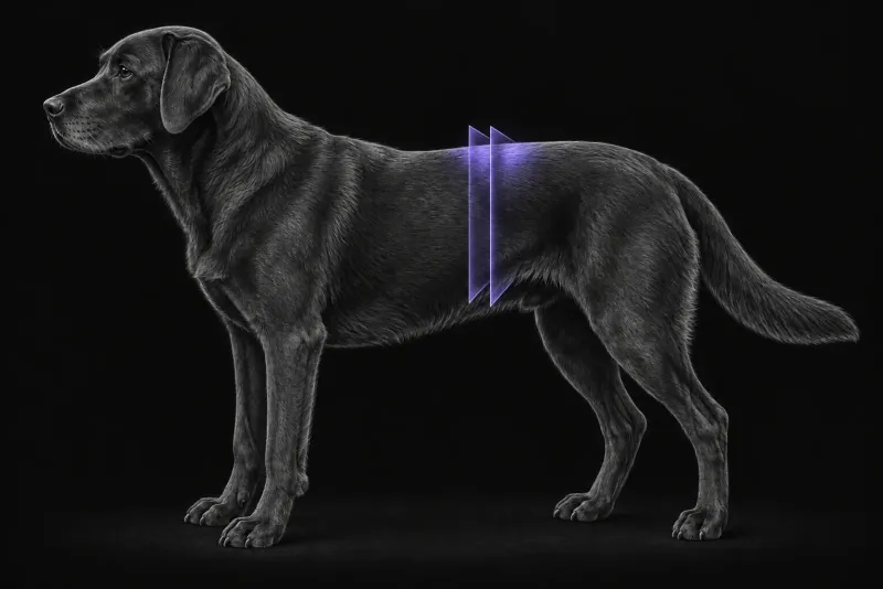
Why adrenal masses need careful imaging
Adrenal masses are often found by chance. In a study of 270 dogs that had abdominal CT for unrelated reasons (Baum et al., Journal of the American Veterinary Medical Association, 2016), 25 (9.3%) had an incidental adrenal mass. These dogs were older (median 12.0 years against 8.2 years). Dogs scanned for neoplasia were more likely to have one (15.9%) than dogs scanned for other reasons (2.3%), although they were also older. Only 3 of the 25 masses were examined histologically.
Once a mass is found, the questions for the surgeon are practical:
- has it entered a vein?
- how far does any tumour thrombus extend?
- which organs is it stuck to?
- what type of tumour is it likely to be?
Image the pituitary too in hypercortisolism
In 201 dogs with spontaneous hypercortisolism that had CT of the pituitary and adrenal glands (van Bokhorst et al., Journal of Veterinary Internal Medicine, 2019), concurrent pituitary and adrenal lesions were present in 5% of all dogs and in 10% of dexamethasone-resistant dogs. The authors recommend imaging both the pituitary and adrenal glands in every dog with spontaneous hypercortisolism.
Vascular invasion: what CT does well
In 15 dogs with 17 adrenal masses (Schultz et al., Veterinary Radiology & Ultrasound, 2009), contrast-enhanced CT identified vascular invasion with 92% sensitivity, 100% specificity and 95% accuracy against surgery or necropsy. In one dog, invasion of the phrenicoabdominal vein was missed. Invasion occurred through the lumen of the phrenicoabdominal vein, with extension inside other veins, rather than by erosion through vessel walls. All four pheochromocytomas in that study invaded nearby vessels.
In 17 dogs with primary adrenal neoplasia (Gregori et al., Veterinary Radiology & Ultrasound, 2015), CT signs of vascular invasion agreed excellently with findings at surgery or necropsy (kappa 0.86). Pheochromocytoma was associated with CT signs of vascular invasion.
The picture is less clear-cut in a larger study. In 91 dogs scored on a 7-point CT scale (Pey et al., Journal of Veterinary Internal Medicine, 2022), a large equivocal zone between scores 3 and 6 lowered the accuracy of CT for vascular invasion. Agreement between radiologists was excellent for the caudal vena cava but good to moderate for other vessels.
The cranial extent of a caval thrombus matters for planning. In 19 dogs that had adrenalectomy with caval venotomy (Knight et al., Veterinary Surgery, 2019), the thrombus reached the prehepatic (14 dogs), hepatic (3) or posthepatic (2) caudal vena cava. The number of vascular tourniquets used reflected where the phrenicoabdominal vein entered the cava and the length of the thrombus. Extension beyond the hepatic hilus did not prevent a good outcome. Perioperative mortality was 21% (4 dogs).
Adhesions to organs and vessels
In 30 dogs and 2 cats that had CT and adrenalectomy (Baldinetti et al., Veterinary Radiology & Ultrasound, 2025), the tumour was adhered to adjacent structures at surgery in 27 of 32 patients (84.4%). No single CT criterion predicted adhesion. Combinations of criteria gave good accuracy for some structures, for example 86.2% for the caudal vena cava with a right-sided tumour and 90% for the kidney.
Tumour type: helpful features, real overlap
| Comparison | CT differences reported |
|---|---|
| Carcinoma vs pheochromocytoma | Differed in pre- and post-contrast attenuation, contralateral adrenal size, size of the tumour thrombus, and mineralisation of tumour and thrombus (7-point scale study). On triple-phase CT, pheochromocytomas had higher maximum attenuation before contrast and in arterial and venous phases, and higher wash-in and washout values (Yoshida et al., Veterinary Surgery, 2016). |
| Adenoma vs carcinoma | Adenomas had a higher minimum pre-contrast value and higher washout between venous and delayed phases (triple-phase CT study). Adenomas and malignant tumours differed in contour irregularity (7-point scale study). |
| Delayed-phase patterns | A peripheral enhancing rim was associated with fibrous encapsulation. A heterogeneous pattern was associated with haemorrhage or infarction (Gregori et al.). |
These features support a probability, not a diagnosis. Overlapping characteristics limited reliable distinction between tumour types on CT alone.
Size and the absence of invasion do not prove a tumour benign either. In 51 dogs with tumours of 3 cm or less and no vascular invasion on CT (Cavalcanti et al., Veterinary and Comparative Oncology, 2020), 28 (54.9%) had a malignancy. Short-term survival after adrenalectomy was 92.2% and one-year disease-specific survival 83.3%.
Adrenal CT protocol at a glance
Adrenal CT depends on a good-quality contrast-enhanced study, because a poor post-contrast study reduced both the performance of CT and agreement between radiologists. Studies of tumour type compared the mass before contrast and in arterial, venous and delayed phases, using triple-phase helical CT. In a dog with hypercortisolism, the pituitary is scanned together with the adrenal glands.
What the radiologist looks at
- Vascular invasion. The caudal vena cava, the phrenicoabdominal vein and other nearby veins, and the cranial extent of any thrombus. Phrenicoabdominal vein invasion can be missed, and agreement is lower for vessels other than the caudal vena cava.
- Adhesions. Contact with the caudal vena cava, kidney and other structures, judged on a combination of CT criteria rather than any single sign.
- Tumour features. Attenuation, enhancement and washout, contour, mineralisation and the size of the other adrenal gland, read as a likely tumour type, not a diagnosis.
- The pituitary, when the dog has hypercortisolism.
What your report will include
Every Veheri case is read by a radiologist or neurologist, and when you order you can choose Diplomate-only reporting or the full specialist panel. You can chat directly with the specialist assigned to your case, and a simplified pet owner report helps you explain the findings to the owner.
When you take the report forward:
- Tumour type still needs histology, because CT features overlap between adenoma, carcinoma and pheochromocytoma.
- A small mass without invasion is not proof of a benign tumour: 54.9% of such tumours were malignant.
- Hormonal activity is a separate question. CT shows anatomy. Whether a tumour is functional is usually answered with endocrine testing.
- Surgical planning depends on the thrombus extent, but extension beyond the hepatic hilus did not prevent a good outcome.
- Talking to the owner: in dogs with small tumours and no vascular invasion, short-term survival after adrenalectomy was 92.2%.
What to include when you request an adrenal CT report
- signalment: age, breed and sex
- clinical signs and endocrine test results, including any dexamethasone suppression results
- whether the mass was an incidental finding
- previous ultrasound findings and measurements, so growth can be judged
- whether adrenalectomy is being considered, so vascular and organ involvement is described in surgical terms
- any previous abdominal surgery
How Veheri can help
Veheri’s radiologists report veterinary CT studies remotely for clinics, and offer expert support with CT protocols, positioning and acquisition. For dogs with hypercortisolism, see pituitary MRI in dogs, and for wider staging, our oncology imaging guide.
Sources
Each claim in this guide links to its source. The full citations:
- Baum JI, Boston SE, Case JB. Prevalence of adrenal gland masses as incidental findings during abdominal computed tomography in dogs: 270 cases (2013-2014). J Am Vet Med Assoc. 2016;249(10):1165-1169. doi:10.2460/javma.249.10.1165
- Schultz RM, Wisner ER, Johnson EG, MacLeod JS. Contrast-enhanced computed tomography as a preoperative indicator of vascular invasion from adrenal masses in dogs. Vet Radiol Ultrasound. 2009;50(6):625-9. doi:10.1111/j.1740-8261.2009.01593.x
- Gregori T, Mantis P, Benigni L, et al. Comparison of computed tomographic and pathologic findings in 17 dogs with primary adrenal neoplasia. Vet Radiol Ultrasound. 2015;56(2):153-9. doi:10.1111/vru.12209
- Pey P, Specchi S, Rossi F, et al. Prediction of vascular invasion using a 7-point scale computed tomography grading system in adrenal tumors in dogs. J Vet Intern Med. 2022;36(2):713-725. doi:10.1111/jvim.16371
- Knight RC, Lamb CR, Brockman DJ, Lipscomb VJ. Variations in surgical technique for adrenalectomy with caudal vena cava venotomy in 19 dogs. Vet Surg. 2019;48(5):751-759. doi:10.1111/vsu.13168
- Baldinetti M, Mattei C, Donato PD, et al. Computed tomography accuracy for prediction of adrenal tumor adhesion with organs and vessels in dogs and cats. Vet Radiol Ultrasound. 2025;66(6):e70096. doi:10.1111/vru.70096
- Yoshida O, Kutara K, Seki M, et al. Preoperative differential diagnosis of canine adrenal tumors using triple-phase helical computed tomography. Vet Surg. 2016;45(4):427-35. doi:10.1111/vsu.12462
- Cavalcanti JVJ, Skinner OT, Mayhew PD, et al. Outcome in dogs undergoing adrenalectomy for small adrenal gland tumours without vascular invasion. Vet Comp Oncol. 2020;18(4):599-606. doi:10.1111/vco.12587
- van Bokhorst KL, Kooistra HS, Boroffka SAEB, Galac S. Concurrent pituitary and adrenocortical lesions on computed tomography imaging in dogs with spontaneous hypercortisolism. J Vet Intern Med. 2019;33(1):72-78. doi:10.1111/jvim.15378
Related reading
More imaging guides
All imaging guides
Brain CT in dogs when MRI is not available
What brain CT can and cannot detect in dogs when MRI is not available, for masses, stroke, inflammation and head trauma, and when MRI or CSF is still needed.
CT of the canine elbow for medial coronoid disease
When CT is worth it for medial coronoid disease in dogs, what it finds that radiographs miss, and where arthroscopy still gives the better answer.
CT of the canine gastrointestinal tract in the acute abdomen
When abdominal CT adds to radiographs and ultrasound in vomiting dogs with suspected obstruction, foreign body or perforation, and what CT can and cannot show.