
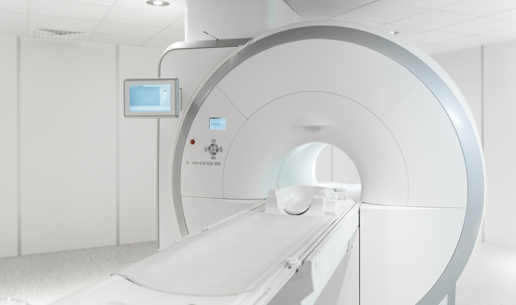
Veterinary CT protocol optimization blends physics, pharmacology, and workflow. Start with task-based protocols, nail motion control, treat contrast like a pharmacologic intervention, and remove unnecessary phases via spectral CT where validated. Use governance frameworks to set culture, and lean on Virtual Scanning Support to lift smaller teams. The payoff is faster, safer scans and sharper decisions across your caseload.
Clinicians searching for“Vet CT Protocol Optimization” want practical, evidence-based settings to improve image quality, reduce dose, and streamline workflows. The short answer: standardize task-based protocols, tailor kVp/mAs and contrast to patient and indication, lock down motion, and use iterative reconstruction and lean workflows.
As a radiology-led writer who has optimized multi-site CT protocols and trained teams across species and scanners, I’ll anchor recommendations in peer-reviewed veterinary literature and trusted protocol governance. You’ll also find realistic tips for small teams, including how to leverage Virtual Scanning Support when expertise is remote.
What “optimization” means in veterinary CT
Optimization in veterinary CT means achieving the diagnostic task with the lowest reasonable dose and the most reliable image quality for the patient’s size, indication, and scanner. It also means reducing repeats, anesthesia time, and handoffs—key wins for busy hospitals. In practice, that translates to standardized protocols per indication, patient-adjusted dose and contrast, aggressive motion control, and data-driven QA.
Build your task-based protocol library
Moving from a generic “chest/abdomen” to task-based protocols is the fastest route to consistent quality and predictable dose. Start with common indications (head, thorax, abdomen, elbow/stifle, CTA, dental/maxillofacial) and pre-define scan range, phases, kVp, mAs or noise index, pitch, rotation time, kernel, and reconstruction thickness. For vascular and arthrographic studies, use current animal data to set initial parameters and tighten them through QA and radiologist feedback. Contemporary canine angio-CT and CTA research offers practical guardrails for timing and intra-articular dosing; build these learnings into your protocol cards and weight bands.
“Computed tomography (CT) imaging is vital for diagnosing and monitoring diseases in both humans and animals, yet radiation exposure remains a significant concern, especially in animal imaging.” — Yuseong Son (Hankuk University of Foreign Studies), inMDPI Biomedicines Engineering
Also Read:Vet Imaging Solutions & Info
Dose governance: set a culture and targets
Dose optimization is as much culture as it is physics. Adopt human-validated governance frameworks and adapt them to species and sizes you serve. Establish Local Diagnostic Reference Levels (LDRLs), audit them regularly, and train technologists on indication-based protocols and iterative reconstruction choices. Use a protocol committee to manage versioning, change control, and performance metrics.
“CT protocols are a facility’s ongoing mechanism of ensuring that exams being performed achieve the desired diagnostic image quality while optimizing radiation dose.” —Dianna D. Cody,American Association of Physicists in Medicine (AAPM)
For modality-specific guidance in dogs, a recent review synthesizes how injection rate, bolus timing (tracking/test bolus), saline flush, and iodine dosing influence vascular and parenchymal enhancement in abdominal CTA. See the summary in MDPI Animals.
Also Read: The Future of Veterinary Radiology
Motion control and anesthesia: plan for the scan you need
Motion ruins the best protocol. For thorax and angiographic studies, use short rotation times, appropriate pitch, and breath-holds where safe. While most small-animal CT requires sedation or brief anesthesia, selected indications can be done awake with purpose-built restraint to avoid anesthetic risk and speed throughput. Plan fallback options if motion appears mid-exam.
Conscious workflows are especially useful for fragile patients and respiratory evaluations. When scanning awake, prefer high-pitch, short scan times, and be mindful of helical artifacts and SNR/CNR trade-offs.
Contrast media: dose, rate, and timing that work in animals
Contrast strategy drives angiography quality more than almost any other choice. In dogs, evidence emphasizes injection rate, robust bolus timing (tracking or test bolus), saline flush, and dosing adjusted for body composition. A systematic review of canine abdominal angio-CT highlights these drivers and notes that higher iodine concentration doesn’t automatically yield better enhancement when other parameters are optimized. See synthesis in MDPI Animals.
Pragmatically, define arterial-phase CTA with short injection durations and real-time tracking; define portal-venous with fixed timing calibrated to table speed and coverage. Build weight-band tables for mgI/kg and mL/s and standardize the saline flush to reduce streaking and improve bolus utilization.
Reduce phases when technology allows
Multi-phase protocols multiply dose and time. Detector-based spectral CT and validated virtual non-contrast (VNC) can remove the need for true unenhanced phases in selected contexts, trimming dose and exam time where equivalence is proven. Recent work in rabbits using spectral detector CT reported that pre-contrast series may be safely omitted in specific scenarios with proportional dose/time benefits; see Frontiers in Veterinary Science.
If your fleet includes spectral CT, validate VNC against your most common indications and pathologies before changing care pathways. Assess organ-specific performance and edge cases with a blinded read and clear acceptance criteria.
Iterative reconstruction(IR) noise targets, and scanners
Modern iterative reconstruction allows lower mAs for similar noise, but species diversity means you should set indication-based noise or diagnostic targets rather than a single mAs recipe. On older scanners, choose kernels and IR strengths that avoid over-smoothing in bone, preserving trabecular detail for ortho studies. On newer vet‑dedicated systems, features such as metal artifact reduction and AI-driven automation directly support dose and motion control; see the veterinary SOMATOM go.Up.
Also Read: CT case report featured in Vet Practice Magazine Australia
Virtual Scanning Support: multiply expertise across sites
When your expert is off‑site, remote protocol supervision and coaching can save a case. Virtual Scanning Support covers live assistance for protocol selection, parameter tweaks, artifact troubleshooting, and training—especially valuable for after‑hours or satellite clinics. Standardized checklists and protocol cards ensure a consistent baseline that remote experts can refine case by case.
“If you really have a shortage of technicians and you have to decide every morning whether you have to close a machine, yes or no, that’s not really convenient. I mean we totally reduced that down to zero,” —Professor Michael Forsting, Director of the Institute of Diagnostic and Interventional Radiology and Neuroradiology at Essen University Hospital
Even without a formal remote platform, structured messaging, saved protocol cards, dose/contrast calculators, and a rigid QA loop will deliver most of the benefit. For formal tooling, ensure privacy, role-based access, and audit which remote changes reduce repeats and anesthesia time.
Quick-start checklist (print for your console)
You can put these into your protocol cards today. The aim is predictable image quality, fewer repeats, and lower total dose.
- Set task-based protocols with clear scan range, kVp, mAs/noise index, pitch, rotation time, kernel, and recon thickness per indication and size tier.
- For CTA: short injection duration, saline flush, bolus tracking, and table speed aligned to patient length; document mgI/kg and mL/s by weight band.
- Audit regularly: repeat rate, anesthesia duration, CTDIvol/DLP by indication/size, and adherence to protocol—then iterate.
FAQs
Q: What’s the single biggest driver of arterial CTA quality in dogs?A: Injection rate and timing; use bolus tracking and a saline flush, and keep the injection duration short.
Q: Can I do awake thoracic CT in cats?A: Yes, with purpose-built restraint and tuned protocols, published data show high completion rates; ensure contingency plans for motion and patient comfort.
Q: How do I decide whether to skip the true non‑contrast series?A: Validate VNC on your scanner for your common indications; only omit true unenhanced when equivalence is demonstrated locally.
Smarter Teleradiology. Faster Diagnosis. Better Results Join the Veheri Community!
Start today: Book your DEMO
Imaging guides on this topic
Browse all imaging guides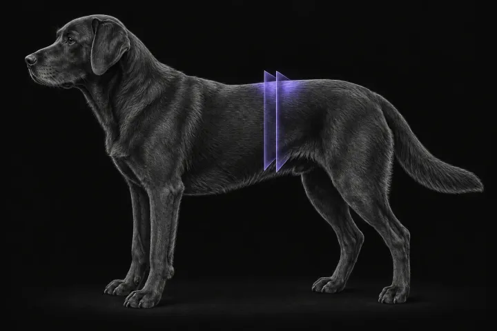
CT of adrenal masses in dogs
What contrast CT shows in a dog with an adrenal mass, from caval invasion and adhesions to tumour type, and where its accuracy runs out before adrenalectomy.
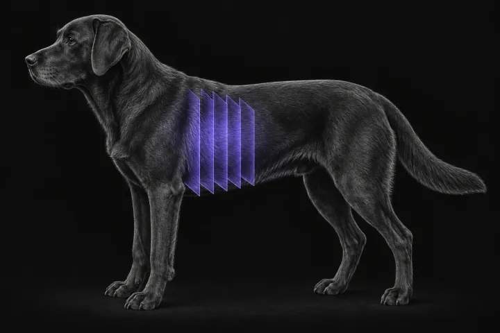
Thoracic CT for pulmonary metastases in dogs
How thoracic CT compares with radiographs for pulmonary metastasis screening in dogs, which tumours most often spread to the lung, and how to read a nodule.
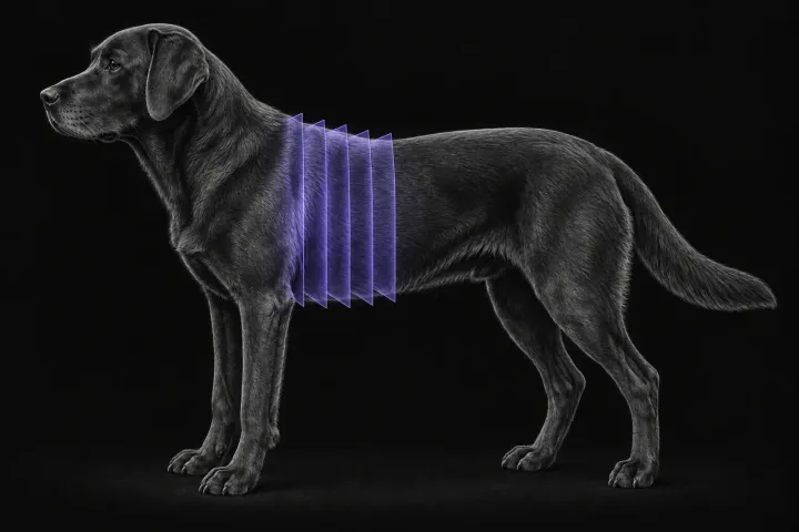
CT pulmonary angiography for suspected pulmonary thromboembolism in dogs
When to request CT pulmonary angiography for suspected pulmonary thromboembolism in dogs, what a positive scan looks like, and why technique decides the answer.