
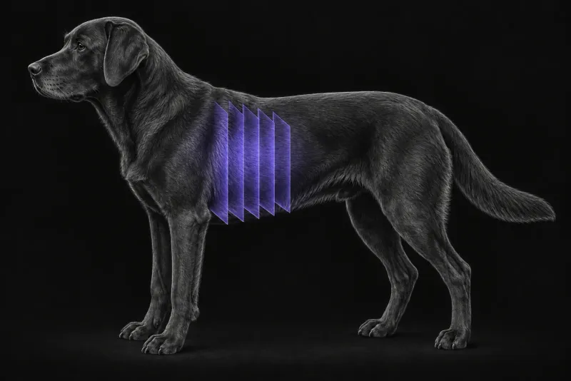
Why lung staging changes decisions
Whether a tumour has already spread to the lungs is usually central to the treatment plan. How likely spread is depends heavily on tumour type. In 536 dogs with non-pulmonary malignant neoplasia that had CT at a single hospital (Lamb et al., Veterinary Radiology & Ultrasound, 2019), multiple pulmonary nodules were present at the first CT in:
| Tumour type | Dogs with multiple nodules at first CT |
|---|---|
| Haemangiosarcoma | 24 of 58 (41%) |
| Osteosarcoma | 14 of 55 (26%) |
| Carcinoma | 20 of 80 (25%) |
| Histiocytic sarcoma | 5 of 21 (24%) |
| Soft tissue sarcoma | 13 of 57 (23%) |
| Adenocarcinoma | 11 of 60 (18%) |
| Melanoma | 5 of 37 (14%) |
| Lymphoma | 10 of 76 (13%) |
| Mast cell tumour | 2 of 47 (4%) |
| Squamous cell carcinoma | 0 of 17 (0%) |
Grade matters too. In 137 dogs with cutaneous or subcutaneous soft tissue sarcoma (Villedieu et al., Journal of the American Veterinary Medical Association, 2021), nodules suggestive of metastasis were found in 6% with grade 1, 6% with grade 2 and 38% with grade 3 tumours. The authors considered lung staging low-yield for grade 1 or 2 tumours, especially those present for 3 months or less.
CT compared with thoracic radiographs
- Size threshold. In 18 dogs with confirmed pulmonary metastases (Nemanic et al., Journal of Veterinary Internal Medicine, 2006), only 9% of the nodules found on CT were visible on radiographs. CT detected nodules down to about 1 mm, while radiographs needed about 7–9 mm to show them reliably. CT also found nodules in more lung lobes.
- Patient level. In 33 dogs with neoplasia (Armbrust et al., Journal of the American Veterinary Medical Association, 2012), CT found nodules or masses in 21. At least one of three radiograph readers saw them in 17 of those 21 dogs. Radiographic sensitivity ranged from 71% to 95% between readers. The 4 dogs missed on radiographs were all large or giant breeds with osteosarcoma, and the authors recommend thoracic CT in these dogs if finding nodules would change treatment.
- Osteosarcoma staging. In a pilot study of 15 dogs with appendicular bone tumours (Oblak et al., Veterinary and Comparative Oncology, 2015), thoracic CT showed lesions that were not visible on radiographs. In 39 dogs staged with whole-body CT (Talbott et al., Veterinary Surgery, 2017), pulmonary metastasis was considered definitive in 2 (5%), and 2 more had soft tissue masses consistent with concurrent, non-metastatic malignancies.
Lung CT protocol at a glance
Metastasis screening needs well-aerated lungs, because collapsed or poorly aerated lung can hide or mimic nodules. In anaesthetised dogs, a fixed positive end-expiratory pressure valve fitted immediately after induction reduced atelectasis (Allison et al., Journal of Small Animal Practice, 2017), and in normal dogs scanned under injectable sedation, no atelectasis was identified (Hunt and Wallack, Veterinary Radiology & Ultrasound, 2021). It is worth agreeing positioning, ventilation and the order of scanning with the anaesthetist before the thorax is imaged.
What the radiologist looks at
CT finds more than radiographs, and not every nodule is a metastasis.
- One nodule or many. A single nodule was seen at first CT in 33 dogs (6%). Of 9 with follow-up CT, the nodule disappeared in 2, did not change in 3, and enlarged with new nodules in 4. The authors call a solitary nodule an indeterminate finding that may be unrelated to the tumour.
- Ground-glass lesions in bone tumours. Equivocal ground-glass lesions progressed in 3 of 4 cases, and the authors advise treating them as suspicious for metastasis.
- Age changes. In a comparison of young and old dogs (Hornby and Lamb, Veterinary Radiology & Ultrasound, 2017), visible foci of heterotopic bone were more common in old dogs (62% vs 14%). Lung collapse was associated with old age, greater body weight and anaesthesia. The authors judged that neither difference should lead to a misdiagnosis of pulmonary disease.
- Atelectasis. Collapsed or poorly aerated lung, which can hide or mimic nodules.
What your report will include
Every Veheri case is read by a radiologist or neurologist, and when you order you can choose Diplomate-only reporting or the full specialist panel. You can chat directly with the specialist assigned to your case, and a simplified pet owner report helps you explain the findings to the owner.
When you take the report forward:
- A solitary nodule that would change the plan: a follow-up CT after an interval is often worth discussing before deciding on one scan, because its behaviour on follow-up varied.
- CT does not give a histological diagnosis. A nodule that does not behave as expected on follow-up still needs sampling or clinical judgement.
- Older radiographs are not a like-for-like baseline, because CT finds smaller nodules than radiographs.
What to include when you request a thoracic CT report
- signalment: age, breed and sex
- tumour type, grade and site, and how long the mass has been present
- whether the result will change treatment
- previous thoracic radiographs or CT, for comparison
- sedation or general anaesthesia, recumbency and ventilation used during the scan
- any known lung disease or previous thoracic surgery
How Veheri can help
Veheri’s radiologists report veterinary CT studies remotely for clinics, and offer expert support with CT protocols, positioning and acquisition. For cats, see thoracic CT in cats, and for wider staging, our oncology imaging guide.
Sources
Each claim in this guide links to its source. The full citations:
- Lamb CR, Whitlock J, Foster-Yeow ATL. Prevalence of pulmonary nodules in dogs with malignant neoplasia as determined by CT. Vet Radiol Ultrasound. 2019;60(3):300-305. doi:10.1111/vru.12723
- Villedieu EJ, Petite AF, Godolphin JD, et al. Prevalence of pulmonary nodules suggestive of metastasis at presentation in dogs with cutaneous or subcutaneous soft tissue sarcoma. J Am Vet Med Assoc. 2021;258(2):179-185. doi:10.2460/javma.258.2.179
- Nemanic S, London CA, Wisner ER. Comparison of thoracic radiographs and single breath-hold helical CT for detection of pulmonary nodules in dogs with metastatic neoplasia. J Vet Intern Med. 2006;20(3):508-15. doi:10.1892/0891-6640(2006)20[508:cotras]2.0.co;2
- Armbrust LJ, Biller DS, Bamford A, et al. Comparison of three-view thoracic radiography and computed tomography for detection of pulmonary nodules in dogs with neoplasia. J Am Vet Med Assoc. 2012;240(9):1088-94. doi:10.2460/javma.240.9.1088
- Oblak ML, Boston SE, Woods JP, et al. Comparison of concurrent imaging modalities for staging of dogs with appendicular primary bone tumours. Vet Comp Oncol. 2015;13(1):28-39. doi:10.1111/vco.12016
- Talbott JL, Boston SE, Milner RJ, et al. Retrospective evaluation of whole body computed tomography for tumor staging in dogs with primary appendicular osteosarcoma. Vet Surg. 2017;46(1):75-80. doi:10.1111/vsu.12579
- Hornby NL, Lamb CR. Does the computed tomographic appearance of the lung differ between young and old dogs? Vet Radiol Ultrasound. 2017;58(6):647-652. doi:10.1111/vru.12532
- Allison A, Huizing X, Jolliffe C, et al. Effect of fixed value positive end expiratory pressure valves on canine thoracic volume and atelectasis. J Small Anim Pract. 2017;58(11):645-651. doi:10.1111/jsap.12710
- Hunt TD, Wallack ST. Minimal atelectasis and poorly aerated lung on thoracic CT images of normal dogs acquired under sedation. Vet Radiol Ultrasound. 2021;62(6):647-656. doi:10.1111/vru.13016
Related reading
More imaging guides
All imaging guides
Thoracic CT in cats for lung tumours, airway and interstitial disease
What thoracic CT adds over radiographs in cats with lung tumours, bronchial or interstitial disease, how anaesthesia affects the lungs, and where CT stops.

CT of adrenal masses in dogs
What contrast CT shows in a dog with an adrenal mass, from caval invasion and adhesions to tumour type, and where its accuracy runs out before adrenalectomy.

Brain CT in dogs when MRI is not available
What brain CT can and cannot detect in dogs when MRI is not available, for masses, stroke, inflammation and head trauma, and when MRI or CSF is still needed.