
Why the mass needs a diagnosis before treatment
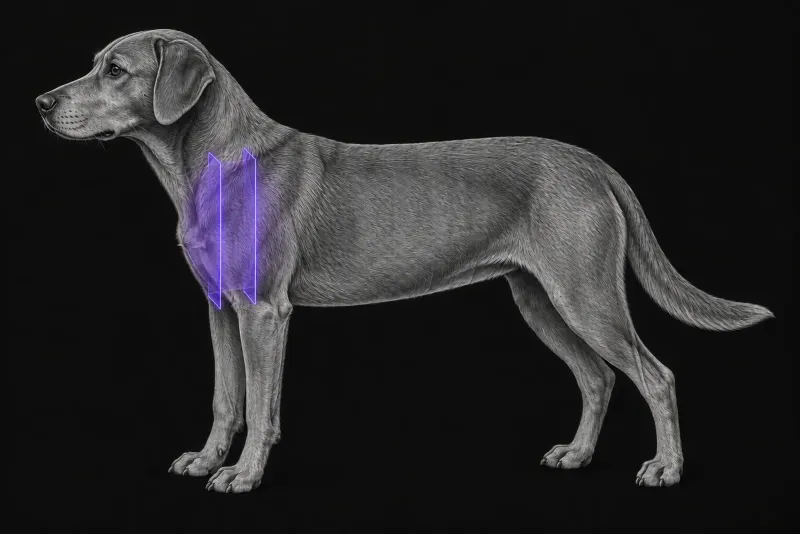
In dogs, the most common cranial mediastinal masses are lymphoma and thymic epithelial tumours (thymoma and thymic carcinoma). The two are treated differently: lymphoma medically, thymic epithelial tumours surgically. Telling them apart before treatment therefore matters, and it needs fine-needle aspiration or biopsy (Oura et al., Journal of the American Animal Hospital Association, 2019).
What radiographs can and cannot say
Radiographs usually find the mass first, but they are limited:
- Origin of the mass. In 75 dogs and cats with masses confirmed on CT (Ruby et al., Veterinary Radiology & Ultrasound, 2020), agreement between radiographs and CT on the origin of the mass was only moderate: 68.6% for mediastinal and 63% for pulmonary masses.
- Type of mass. In 62 dogs, a mass with at least two well-defined margins on the lateral view, or a rightward shift of the cardiac silhouette on the ventrodorsal or dorsoventral view, was more likely to be a thymic epithelial tumour than lymphoma. Radiographic findings did not remove the need for sampling.
What CT shows: thymic epithelial tumour or lymphoma
In a case-control study of 62 dogs with thoracic CT and a confirmed diagnosis (Reeve et al., Veterinary Radiology & Ultrasound, 2020) (33 lymphomas, 24 thymomas and 5 thymic carcinomas), several CT signs differed between the groups:
| Finding on CT | Thymic epithelial tumour | Lymphoma |
|---|---|---|
| Attenuation before and after contrast | Heterogeneous (odds ratio 23.3 pre-contrast, 30.7 post-contrast) | More homogeneous |
| Cranial vena cava | Less likely to be enveloped (odds ratio 0.07) | More likely to be enveloped |
| Standard deviation of HU in the mass after contrast | Greater. Above 17 HU: sensitivity 72%, specificity 79% for thymic epithelial tumour | Smaller |
| Median age | 8.6 years | 6.0 years |
There were no significant differences in morphology, calcification, mediastinal lymphadenopathy, cranial vena cava invasion, collateral vessels or pleural fluid.
Not every cranial mediastinal finding is a tumour. In 866 canine thoracic CT studies (Nanni et al., Veterinary Radiology & Ultrasound, 2024), presumed mediastinal cysts were found in 5.66%. They were mostly small, round, solitary, fluid-attenuating lesions in the cranioventral mediastinum, with well-defined margins, homogeneous attenuation and no contrast enhancement.
Invasion, size and the surgical approach
When the mass is a thymoma, CT describes what the surgeon will meet. In 22 dogs with confirmed thymoma (von Stade et al., Veterinary Radiology & Ultrasound, 2019):
- 19 of 22 masses enhanced heterogeneously
- vascular invasion was present in 7 of 22 dogs, and larger tumours were associated with vascular invasion; larger tumours were also more likely to be cystic
- in fully resected masses, greater tumour height was associated with recurrence
- six dogs had lymphadenopathy on CT, and five of these six had no evidence of metastasis
Tumour size on CT was also related to conversion during thoracoscopic surgery. In 49 dogs that had thoracoscopic removal of a cranial mediastinal mass (Carroll et al., Journal of the American Veterinary Medical Association, 2024), the median maximal tumour diameter on CT was 4.7 cm (range 2.7 to 8.5 cm). Conversion to an open procedure was needed in 4 of 49 dogs (8%), and these dogs had a significantly larger median maximal tumour diameter on CT. Thymoma was the most common tumour (37 of 49).
Outcome after surgery depends on more than imaging. In 80 dogs that had a thymic epithelial tumour excised (Garneau et al., Veterinary Surgery, 2015), perioperative mortality was 20%. Paraneoplastic syndromes and incomplete histological margins were independently associated with shorter survival. In the thoracoscopic series, 17 of 49 dogs (35%) had myasthenia gravis and 11 of 49 (22%) had megaoesophagus before surgery.
Mediastinal CT protocol at a glance
A mediastinal mass is assessed before and after contrast, because attenuation in both phases helped separate thymic epithelial tumours from lymphoma. In thymomas, delayed-phase images were best for judging the degree of enhancement.
What the radiologist looks at
- Signs that shift the probability. Heterogeneous attenuation and a greater spread of HU values favour a thymic epithelial tumour, and envelopment of the cranial vena cava favours lymphoma. These signs do not replace a cytological or histological diagnosis.
- Cyst or tumour. A small, round, fluid-attenuating, non-enhancing lesion fits a presumed mediastinal cyst.
- Invasion and size. Vascular invasion, tumour height and maximal diameter matter for recurrence and for the surgical approach.
- Lymph nodes. Enlarged nodes on CT did not always mean metastasis.
- Oesophagus and lungs. Because myasthenia gravis and megaoesophagus are common in these dogs, it is worth asking for comment on the oesophagus and on the lungs for signs that could fit aspiration.
What your report will include
Every Veheri case is read by a radiologist or neurologist, and when you order you can choose Diplomate-only reporting or the full specialist panel. You can chat directly with the specialist assigned to your case, and a simplified pet owner report helps you explain the findings to the owner.
When you take the report forward:
- Sampling still decides. CT can guide where to sample, but the diagnosis comes from cells or tissue. Both lymphoma and thymoma can consist of small lymphocytes, so cytology of an aspirate can be inconclusive (Lana et al., Journal of Veterinary Internal Medicine, 2006).
- Flow cytometry can help. In that study, aspirates from all six thymomas had at least 10% of lymphocytes co-expressing CD4 and CD8, against less than 2% in six of seven lymphomas. The remaining lymphoma could be distinguished by light scatter, so all 13 masses were identified.
- Surgical planning uses the reported size and invasion, since larger tumours were linked to conversion to open surgery.
- Talking to the owner: outcome after surgery also depends on paraneoplastic syndromes and incomplete margins, not on imaging alone.
What to include when you request a mediastinal CT report
- signalment: age, breed and sex
- the thoracic radiographs and the question you want answered (origin, invasion, resectability or staging)
- signs of myasthenia gravis, regurgitation or megaoesophagus
- calcium and other paraneoplastic findings
- cytology or flow cytometry results, if sampling has already been done
- whether thoracoscopic or open surgery is being considered
How Veheri can help
Veheri’s radiologists report veterinary CT studies remotely for clinics. For cats, see CT of the feline mediastinum, and for staging, read our practical guide to oncology imaging.
Sources
Each claim in this guide links to its source. The full citations:
- Oura TJ, Hamel PE, Jennings SH, et al. Radiographic differentiation of cranial mediastinal lymphomas from thymic epithelial tumors in dogs and cats. J Am Anim Hosp Assoc. 2019;55(4):187-193. doi:10.5326/JAAHA-MS-6907
- Ruby J, Secrest S, Sharma A. Radiographic differentiation of mediastinal versus pulmonary masses in dogs and cats can be challenging. Vet Radiol Ultrasound. 2020;61(4):385-393. doi:10.1111/vru.12859
- Reeve EJ, Mapletoft EK, Schiborra F, et al. Mediastinal lymphoma in dogs is homogeneous compared to thymic epithelial neoplasia and is more likely to envelop the cranial vena cava in CT images. Vet Radiol Ultrasound. 2020;61(1):25-32. doi:10.1111/vru.12812
- Nanni C, Quinci M, Frondizi N, et al. Presumed mediastinal cysts have a low prevalence in canine thoracic computed tomographic studies. Vet Radiol Ultrasound. 2024;65(2):99-106. doi:10.1111/vru.13328
- von Stade L, Randall EK, Rao S, et al. CT imaging features of canine thymomas. Vet Radiol Ultrasound. 2019;60(6):659-667. doi:10.1111/vru.12798
- Carroll KA, Mayhew PD, Culp WTN, et al. Thoracoscopic removal of cranial mediastinal masses in dogs is associated with a low conversion rate, excellent survival to discharge, and good long-term outcome. J Am Vet Med Assoc. 2024;262(10):1-8. doi:10.2460/javma.23.12.0679
- Garneau MS, Price LL, Withrow SJ, et al. Perioperative mortality and long-term survival in 80 dogs and 32 cats undergoing excision of thymic epithelial tumors. Vet Surg. 2015;44(5):557-64. doi:10.1111/j.1532-950X.2014.12304.x
- Lana S, Plaza S, Hampe K, et al. Diagnosis of mediastinal masses in dogs by flow cytometry. J Vet Intern Med. 2006;20(5):1161-5. doi:10.1892/0891-6640(2006)20[1161:dommid]2.0.co;2
Related reading
More imaging guides
All imaging guides
CT of the feline mediastinum for a cranial mediastinal mass
What CT adds for a cat with a cranial mediastinal mass, how far it separates thymoma, lymphoma and cysts, and why cytology or histology still decides.

CT of adrenal masses in dogs
What contrast CT shows in a dog with an adrenal mass, from caval invasion and adhesions to tumour type, and where its accuracy runs out before adrenalectomy.

Brain CT in dogs when MRI is not available
What brain CT can and cannot detect in dogs when MRI is not available, for masses, stroke, inflammation and head trauma, and when MRI or CSF is still needed.