
Why the diagnosis matters
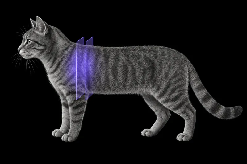
Thymoma and mediastinal lymphoma are the most common cranial mediastinal masses in cats. They differ in treatment and prognosis, so an accurate diagnosis before treatment is essential (Goda et al., Canadian Veterinary Journal, 2026). Benign mediastinal cysts (Camero et al., Journal of Feline Medicine and Surgery) are a third important differential.
The three conditions are described in different groups of cats:
- Mediastinal lymphoma. In 55 cats from five UK referral centres (Fabrizio et al., Journal of Feline Medicine and Surgery), the median age was 3 years (range 0.5 to 12 years), 21.8% were Siamese and the male to female ratio was 3.2:1. Only 5 cats were FeLV-positive.
- Thymic epithelial tumours. In 64 cats (Marks et al., Journal of Small Animal Practice), paraneoplastic syndromes were present in 9. Median tumour diameter was 6 cm (range 2 to 15 cm), and a cystic appearance was described on imaging in 25 cats.
- Mediastinal cysts. In six cats, cysts were incidental findings in cats examined for non-thoracic disease. The cysts recurred in five cats but were never associated with clinical signs.
From the radiograph to CT
A lateral radiograph often raises the question first. In 76 cats (Moore et al., Journal of the American Veterinary Medical Association, 2025), the position of the tracheal bifurcation on a right lateral view helped. A distance of 7.5 cm or more from the caudal manubrium to the bifurcation, and/or 6 cm or more from the first rib to the bifurcation, indicated a possible mediastinal mass and warranted further diagnostics.
Radiographs show that a mass is present. CT adds the local picture. In a series of 14 dogs and cats with histologically confirmed mediastinal malignancies (Yoon et al., Veterinary Radiology & Ultrasound, 2004), CT provided reasonably accurate local staging information.
Can CT tell thymoma from lymphoma?
Not reliably, on current evidence. The studies are small.
- In the 14-patient series, there was no clinically exploitable relationship between the CT appearance and the histological type of the mass.
- A 2026 pilot study of six cats with triple-phase contrast-enhanced CT described different enhancement behaviour:
| Triple-phase CT feature | Thymoma | Mediastinal lymphoma |
|---|---|---|
| Timing of enhancement | Peak in the equilibrium phase | Rapid enhancement in the arterial phase, peak in the venous phase |
| Distribution of contrast | Heterogeneous | Homogeneous |
The authors stress the small sample size and that the study was designed to propose a hypothesis that larger studies must test.
Cysts. In the one cat of the cyst series that had CT, the cyst appeared as a well-defined, fluid-attenuating mass in the cranial mediastinum without peripheral contrast enhancement. Thymic epithelial tumours can also look cystic on imaging, so a cystic component on its own does not settle the diagnosis.
What CT detail changes for surgery and prognosis
- Stage of thymic epithelial tumours. In the 64-cat series, surgical excision was attempted in 54 cats, with a perioperative mortality of 11%. Cats with Masaoka-Koga stage I and II tumours survived longer than those with stage III and IV (1366 versus 454 days). Stage was the only significant prognostic factor on multivariable analysis. Recurrence occurred in 11 cats at a median of 564 days.
- Size and surgical approach. In 17 cats that underwent thoracoscopic resection (Renier et al., Veterinary Surgery, 2025), 6 needed conversion to an open or video-assisted procedure. Conversions occurred mainly with masses larger than 5 cm, and larger size and adhesions were associated with conversion. Thymoma was the tumour type in 12 of the 17 cats.
- Lymphoma. In 38 treated cats with mediastinal lymphoma, the overall response to chemotherapy was 94.7% and median survival was 373 days. Cats with a complete response survived longer than those with a partial response (980 versus 42 days).
Mediastinal CT protocol at a glance
The mass is assessed on contrast-enhanced thoracic CT. The 14-patient series assessed contrast enhancement, internal architecture, size, local invasion, pleural fluid and vascular invasion. Triple-phase imaging was used in the six-cat pilot study above.
What the radiologist looks at
- Enhancement pattern. A pattern can favour thymoma or lymphoma, but on current evidence it is usually not a substitute for sampling.
- Solid and fluid parts. These are worth describing, because they help decide where to aim the needle.
- Size and neighbours. For the surgeon, the report should usually state the maximum diameter of the mass, its relation to the heart and great vessels, and any sign of invasion of neighbouring structures. Size above 5 cm was linked to conversion during thoracoscopy.
What your report will include
Every Veheri case is read by a radiologist or neurologist, and when you order you can choose Diplomate-only reporting or the full specialist panel. You can chat directly with the specialist assigned to your case, and a simplified pet owner report helps you explain the findings to the owner.
When you take the report forward:
- Sampling still decides. Fine-needle aspiration can be inconclusive. Even so, in 58 mediastinal masses from 21 dogs and 37 cats (Pintore et al., Journal of Small Animal Practice), agreement between cytological and histological classification ranged from substantial (κ = 0.72) to almost perfect (κ = 0.89), depending on how suspicious cytology results were counted.
- Suspected cysts. In five of the six cats with cysts, aspiration yielded acellular fluid, and afterwards the masses were no longer visible on ultrasound or radiographs.
- Talking to the owner: for thymic epithelial tumours, stage was the only significant prognostic factor, and for lymphoma, cats with a complete response to chemotherapy survived longer than those with a partial response.
What to include when you request a mediastinal CT report
- signalment: age, breed and sex
- FeLV and FIV status
- clinical signs, and any suspected paraneoplastic syndrome
- pleural effusion: whether it was drained, and fluid analysis results
- previous radiographs, ultrasound and cytology results
- previous treatment, including corticosteroids
- whether surgery, chemotherapy or both are being considered, so the report answers the right question
How Veheri can help
Veheri’s radiologists report veterinary CT studies remotely for clinics. For dogs, see CT of the canine cranial mediastinum, and for staging, read our practical guide to oncology imaging.
Sources
Each claim in this guide links to its source. The full citations:
- Goda Y, Hanazono K, Hori A, et al. Triple-phase contrast-enhanced computed tomography findings for feline cranial mediastinal tumors: Pilot study. Can Vet J. 2026;67(3):261-266. PMID: 41847482. https://pubmed.ncbi.nlm.nih.gov/41847482/
- Camero CM, Neumann ZL, Garrett LD. Long-term survival in six cats with mediastinal cysts. J Feline Med Surg. 21(10):998-1002 (published online 2018). doi:10.1177/1098612X18806710
- Fabrizio F, Calam AE, Dobson JM, et al. Feline mediastinal lymphoma: a retrospective study of signalment, retroviral status, response to chemotherapy and prognostic indicators. J Feline Med Surg. 16(8):637-44 (published online 2013). doi:10.1177/1098612X13516621
- Marks TA, Rossanese M, Yale AD, et al. Prognostic factors and outcome in cats with thymic epithelial tumours: 64 cases (1999-2021). J Small Anim Pract. 65(1):47-55 (published online 2023). doi:10.1111/jsap.13675
- Moore AB, Nafe LA, DeAngelis KM, et al. Right lateral thoracic radiographic metrics help discriminate cranial mediastinal masses and pleural effusion in cats. J Am Vet Med Assoc. 2025;263(10):1245-1251. doi:10.2460/javma.24.12.0814
- Yoon J, Feeney DA, Cronk DE, et al. Computed tomographic evaluation of canine and feline mediastinal masses in 14 patients. Vet Radiol Ultrasound. 2004;45(6):542-6. doi:10.1111/j.1740-8261.2004.04093.x
- Pintore L, Bertazzolo W, Bonfanti U, et al. Cytological and histological correlation in diagnosing feline and canine mediastinal masses. J Small Anim Pract. 55(1):28-32 (published online 2013). doi:10.1111/jsap.12161
- Renier S, Massari F, Jimenez Pelaez M, et al. Thoracoscopic resection of cranial mediastinal masses: Retrospective study in 17 cats. Vet Surg. 2025;54(8):1582-1591. doi:10.1111/vsu.70027
Related reading
More imaging guides
All imaging guides
CT of the canine cranial mediastinum
How CT helps separate thymic epithelial tumours from lymphoma in dogs with a cranial mediastinal mass, and what it shows about invasion and surgical approach.
CT of the feline interscapular region for injection-site sarcoma
What contrast CT shows in feline injection-site sarcoma, how it changes surgical and radiotherapy planning, and where imaging cannot replace histology.
Thoracic CT in cats for lung tumours, airway and interstitial disease
What thoracic CT adds over radiographs in cats with lung tumours, bronchial or interstitial disease, how anaesthesia affects the lungs, and where CT stops.