
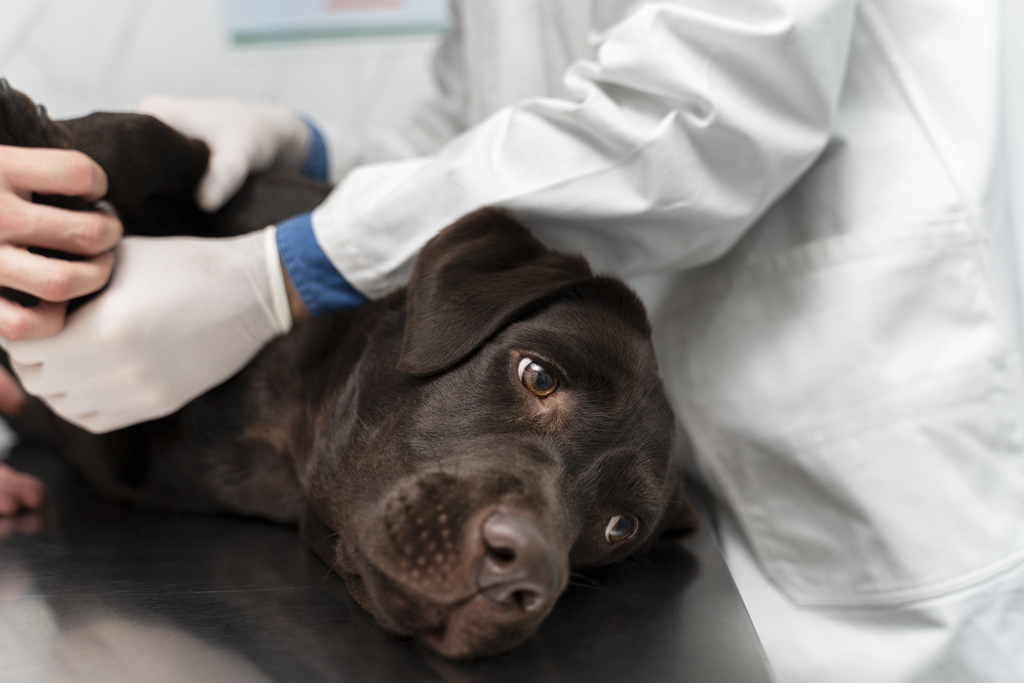
Use radiographs and ultrasound for baseline staging; add CT for local bony detail and sensitive lung metastasis detection; choose MRI for brain and soft-tissue clarity; reserve PET/CT for advanced or research settings. Always integrate node sampling with imaging. This decision-first approach, grounded in peer‑reviewed reviews and major hospital guidance, aligns with how clinicians make time‑sensitive, anesthesia‑aware staging choices.
Veterinary oncology imaging: a practical guide to staging and planning If you’re here to decide “which imaging test when,” start with this: radiographs and ultrasound for baseline staging; CT for local extent and lung metastases; MRI for brain/soft tissue detail; PET/CT is emerging for select cases and trials.
Veterinary oncology imaging underpins diagnosis, staging, prognosis, and treatment planning. This guide distills modality choices by task—local, regional, and distant disease—so you can match the test to the tumor and question. It synthesizes peer‑reviewed reviews and leading hospital guidelines into a practical, decision-first approach.
The role of imaging in cancer care
Imaging confirms where disease is, how far it has spread, and whether surgery, radiation, or systemic therapy is feasible. In practice, you will blend modality strengths with patient stability, anesthesia risk, and access.
- Radiographs/ultrasound: fast, accessible baseline for thorax/abdomen.
- CT: bone and lung detail, 3D planning for surgery/radiation.
- MRI: superior soft tissue/neurologic delineation.
- PET/CT: research/advanced referral cases for functional assessment.
“Imaging is critical for the diagnosis and staging of veterinary oncology patients.” —Katherine S. Hansen & Michael S. Kent,Frontiers in Veterinary Science
Local staging: choosing between CT and MRI
Local staging asks: can I cut it out, irradiate it precisely, or must I palliate? CT excels for bone and complex anatomy (oral/nasal, axial skeleton) and provides the dataset needed for radiation planning. MRI is preferred for brain, spinal cord, and soft-tissue planes where contrast resolution matters most.
- Oral/nasal tumors: CT for bony margins, maxillofacial planning; MRI to assess soft-tissue and intracranial extension when suspected.
- Brain/spine: MRI to map tumor boundaries, edema, and neural involvement.
- Limb tumors: CT clarifies cortical lysis; MRI for intramedullary extent.
Also Read: Diagnostic Imaging Case of the month (Veheri)
Regional nodes: don’t just look—sample
Regional staging asks if lymph nodes are involved. Size and shape on CT/ultrasound can mislead, so plan to aspirate even normal-appearing nodes when it changes management. Consider sentinel lymph node mapping (contrast CT/CEUS/lymphoscintigraphy) for tumors with unpredictable drainage.
- Mandibular/medial retropharyngeal nodes in oral tumors: image all likely basins; aspirate suspicious and “normal” nodes if feasible.
- Axillary/inguinal in limb or trunk tumors: ultrasound/CT for mapping, image-guided FNA.
Distant metastasis: thoracic radiographs vs CT vs PET/CT
Metastatic screening shapes prognosis and treatment choices. Three‑view thoracic radiographs are a baseline standard, but CT is more sensitive for small nodules and can change surgical or radiation plans. PET/CT is reserved for advanced centers and trials.
- Use 3-view thoracic radiographs at minimum for most solid tumors.
- CT chest is preferred when finding small metastases will alter management (e.g., osteosarcoma pre‑amputation, high‑grade sarcomas, oral melanomas).
- PET/CT can reveal functional disease extent beyond contrast CT in select cases.
“Pulmonary metastasis may be revealed by thoracic imaging with 3‑view radiography or CT. These staging tests are important for assessment of prognosis, especially when surgery is being considered.” — Arata Matsuyama,Merck Veterinary Manual
Putting it together: a quick imaging triage
Every case starts with a question: local operability, nodal status, or distant spread. From there, sequence studies to minimize anesthesia episodes and maximize decision value.
- Suspected oral/nasal tumor: head/neck contrast CT for local staging and nodes; thoracic CT if results will alter surgery/radiation; otherwise 3‑view radiographs. Consider MRI if neurologic extension suspected.
- Soft tissue sarcoma on limb/trunk: ultrasound for regional nodes with FNA; thoracic radiographs; CT if surgical margins are complex or met risk high.
- CNS mass: MRI brain/spine; thoracic radiographs or CT if surgery/radiation planned.
- GI mass: abdominal ultrasound (FNA where safe), consider CT for surgical planning and to assess lungs if surgery is planned.
Practicalities: anesthesia, access, and where PET/CT fits
Advanced imaging often needs anesthesia; plan combined protocols to answer multiple questions (e.g., CT head/neck/chest in one setting when it will change management). Not every case needs PET/CT; use it when functional characterization would meaningfully shift therapy, typically in referral/research settings.
“The blueprint has begun to shift where investigations of novel cancer imaging technologies in companion species precede human application and are involved earlier in the development pipeline.” — David Vail, Frontiers in Oncology
Smarter Teleradiology. Faster Diagnosis. Better Results Join the Veheri Community! Start today: Book your DEMO
Imaging guides on this topic
Browse all imaging guides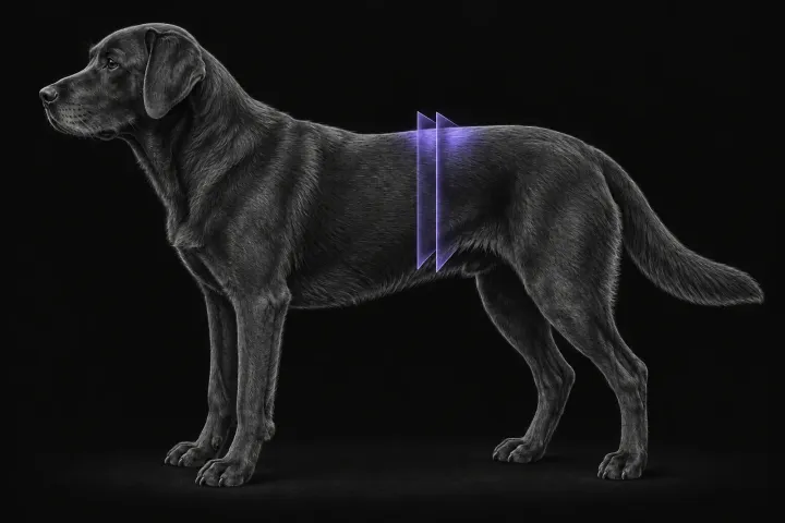
CT of adrenal masses in dogs
What contrast CT shows in a dog with an adrenal mass, from caval invasion and adhesions to tumour type, and where its accuracy runs out before adrenalectomy.
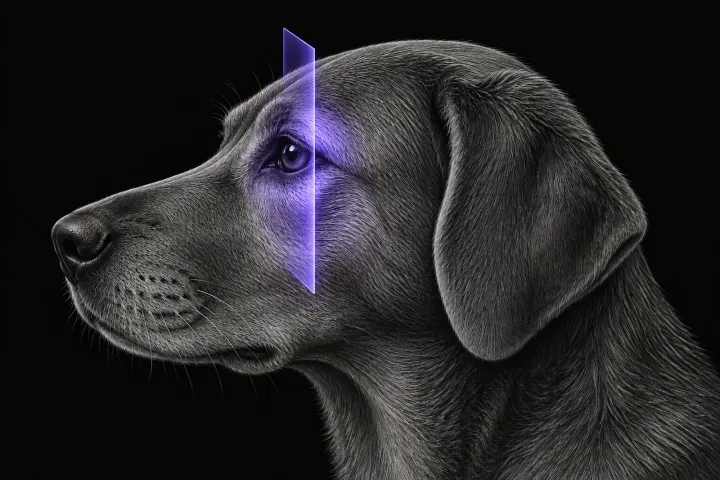
CT of the canine orbit for exophthalmos and retrobulbar disease
How CT helps separate orbital neoplasia from cellulitis and other inflammation in dogs with exophthalmos, where MRI adds detail, and how CT guides sampling.
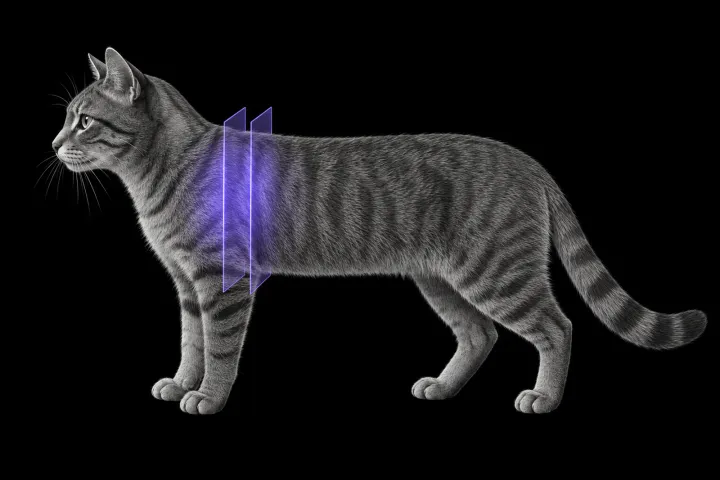
CT of the feline mediastinum for a cranial mediastinal mass
What CT adds for a cat with a cranial mediastinal mass, how far it separates thymoma, lymphoma and cysts, and why cytology or histology still decides.