
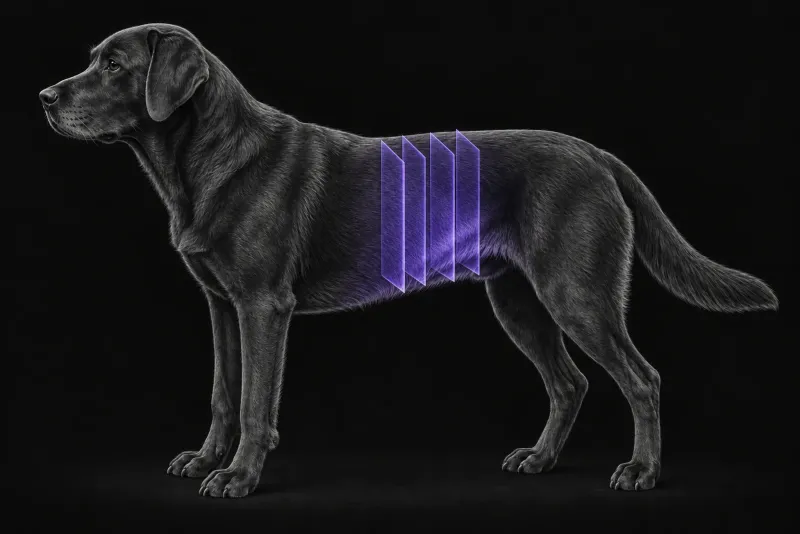
Why the exact anatomy matters
Ureteral ectopia in juveniles and urethral sphincter mechanism incompetence in adults are the two leading causes of urinary incontinence in dogs (Owen, Journal of Small Animal Practice, 2019). Diagnostic accuracy has improved with wider use of CT and cystoscopy. Ectopic ureters are the most common cause of incontinence in young dogs, but should be considered in any incontinent dog with an unknown history (Davidson and Westropp, Veterinary Clinics of North America: Small Animal Practice, 2014).
Treatment depends on the course of the ureter. In 31 female dogs (Hooi et al., Journal of Veterinary Internal Medicine, 2022), cystoscopic-guided laser ablation of intramural ectopic ureters left 67.7% continent, with or without added medical management. The authors note that extramural ectopic ureters need surgical management. The imaging question is therefore not only “ectopic or not”, but also “intramural or extramural, and where does it open”.
Ultrasound, CT and cystoscopy compared
Ectopic ureters can be diagnosed with several techniques, alone or in combination:
- excretory urography
- fluoroscopic urethrography or ureterography
- ultrasound
- cystoscopy
- helical CT
The comparison below uses cystoscopy or a confirmed final diagnosis as the reference. The prospective CTEU study (Schwarz et al., Veterinary Radiology & Ultrasound, 2021) included 42 normal, 27 intramural ectopic and only 3 extramural ectopic ureters.
| Test | What the evidence shows |
|---|---|
| Ultrasound | In 38 incontinent dogs (Taylor et al., Veterinary Radiology & Ultrasound, 2022), sensitivity 93.5% and specificity 100% per dog, against cystoscopy. Per ureter, 87.8% and 86.7%. Performed by an experienced ultrasonographer; not to be relied on alone for individual ureters. |
| CT excretory urography (CTEU) | In 36 dogs, sensitivity 73% and specificity 90.2%. |
| Time-resolved (4D) CTEU | In the same dogs, sensitivity 97% and specificity 94.6%, with nearly perfect agreement between observers. |
| Cystoscopy | The reference standard in these studies. Also the route for laser ablation of intramural ectopic ureters. |
Where CTEU falls short
- CT conclusively and correctly identified 13 of 26 normal ureters (50%) and 32 of 41 ectopic ureters (78%).
- Sensitivity for an extramural course was 29%, and for an intramural course 65%. Specificity was 97% and 71%.
- Sensitivity for the location of the ectopic orifice varied from 0% to 76% depending on the site, and was low especially in or near the urethral sphincter.
The authors conclude that CT findings may need confirmation by cystoscopy, and possibly intraoperative fluoroscopy, before deciding on laser ablation.
flowchart TD
  accTitle: How does the ureter's course change treatment?
  accDescr: From the laser ablation study and the CT and cystoscopy comparison. An intramural ectopic ureter can be treated by cystoscopic-guided laser ablation, after cystoscopy confirms the CT findings where needed. An extramural ectopic ureter needs surgical management.
  start["Ectopic ureter on CTEU"] --> course{{"Intramural or extramural?"}}
  course -- "Intramural" --> confirm["Confirm at cystoscopy if needed"]
  confirm --> laser["Cystoscopic-guided laser ablation"]
course -- "Extramural" --> surgery["Surgical management"]CTEU protocol at a glance
CTEU includes the kidneys and ureters in the same study. In the prospective study, time-resolved (4D) CTEU was more accurate than standard CTEU, and tilting the pelvis with a wedge did not increase confidence about the ureteral opening. In 24 dogs (Secrest et al., Veterinary Radiology & Ultrasound, 2017), thin-slab maximum intensity projection reconstructions increased reader confidence.
What the radiologist looks at
- Signs of an intramural ectopic ureter. In 10 dogs with 14 confirmed intramural ectopic ureters (Fox et al., Journal of Small Animal Practice, 2016), reviewers identified all 14. Every one lacked a normal ureterovesical junction, had a urethral orifice and lacked ureteral divergence. Ureteral dilatation was seen in 7 and tortuosity in 3.
- The ureterovesical junction angle. An obtuse angle was significantly more common in intramural ectopic than in normal ureters and increased diagnostic confidence.
- Reconstructions. When read by a radiologist, thin-slab maximum intensity projection images were similar to or better than transverse source images in accuracy.
- The rest of the urinary tract. Dogs with ectopic ureters can have renal agenesis or dysplasia, hydronephrosis, hydroureter and vestibulovaginal anomalies.
- Each ureter separately. Normal or ectopic; for each ectopic ureter, whether the course is intramural or extramural, where the orifice appears to be, and how certain each call is.
What your report will include
Every Veheri case is read by a radiologist or neurologist, and when you order you can choose Diplomate-only reporting or the full specialist panel. You can chat directly with the specialist assigned to your case, and a simplified pet owner report helps you explain the findings to the owner.
When you take the report forward:
- A stated uncertainty is useful. A report that states its own confidence is usually more useful to the surgeon than one that forces a call. The cystoscopist can then check the doubtful ureter directly, as the CT and cystoscopy comparison advises.
- An extramural course points to surgical management rather than laser ablation.
- Talking to the owner: after laser ablation of intramural ectopic ureters, 67.7% of 31 female dogs were continent, with or without added medical management.
What to include when you request a CTEU report
- signalment: age, breed and sex
- whether incontinence has been present since birth
- ultrasound findings, including any upper urinary tract changes
- urine culture results and previous treatment
- any previous ureteral surgery or laser ablation
- whether cystoscopy or laser ablation is planned
How Veheri can help
Veheri provides veterinary teleradiology for X-ray, CT and MRI, with expert support for CT scanning protocols, positioning and acquisition. Related reading: CT of the feline ureters for suspected obstruction, CT of the canine kidneys and why the quality of the clinical history matters.
Sources
Each claim in this guide links to its source. The full citations:
- Owen LJ. Ureteral ectopia and urethral sphincter mechanism incompetence: an update on diagnosis and management options. J Small Anim Pract. 2019;60(1):3-17. doi:10.1111/jsap.12950
- Davidson AP, Westropp JL. Diagnosis and management of urinary ectopia. Vet Clin North Am Small Anim Pract. 2014;44(2):343-53. doi:10.1016/j.cvsm.2013.11.007
- Hooi KS, Vachon C, Martel DV, et al. Retrospective evaluation of cystoscopic-guided laser ablation of intramural ectopic ureters in female dogs. J Vet Intern Med. 2022;36(1):156-163. doi:10.1111/jvim.16321
- Schwarz T, Bommer N, Parys M, et al. Four-dimensional CT excretory urography is an accurate technique for diagnosis of canine ureteral ectopia. Vet Radiol Ultrasound. 2021;62(2):190-198. doi:10.1111/vru.12934
- Taylor O, Knight R, Genain MA, et al. Ultrasonography as a sensitive and specific diagnostic modality for the detection of ectopic ureters in urinary incontinent dogs. Vet Radiol Ultrasound. 2022;63(3):328-336. doi:10.1111/vru.13055
- Song MK, Fracka AB, Karn K, et al. Receiver operating characteristics of computed tomography (CT) compared to cystoscopy in diagnosis of canine ectopic ureters: thirty-five cases. Vet Surg. 2024;53(3):494-502. doi:10.1111/vsu.14064
- Fox AJ, Sharma A, Secrest SA. Computed tomographic excretory urography features of intramural ectopic ureters in 10 dogs. J Small Anim Pract. 2016;57(4):210-3. doi:10.1111/jsap.12460
- Secrest S, Bugbee A, Waller K, et al. Comparison of transverse computed tomographic excretory urography images and maximum intensity projection images for diagnosing ectopic ureters in dogs. Vet Radiol Ultrasound. 2017;58(2):163-168. doi:10.1111/vru.12461
Related reading
More imaging guides
All imaging guides
CT of the feline ureters for suspected ureteral obstruction
How non-enhanced CT compares with ultrasound for ureteroliths in cats, why calculus count and position matter, and what CT cannot show.

CT of adrenal masses in dogs
What contrast CT shows in a dog with an adrenal mass, from caval invasion and adhesions to tumour type, and where its accuracy runs out before adrenalectomy.

Brain CT in dogs when MRI is not available
What brain CT can and cannot detect in dogs when MRI is not available, for masses, stroke, inflammation and head trauma, and when MRI or CSF is still needed.