
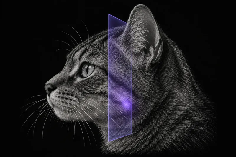
Why the middle ear is worth imaging in cats
Middle ear disease is a common cause of vestibular signs in cats. In 196 cats with peripheral vestibular syndrome that all had CT or MRI of the head (Caldero Carrete et al., Veterinary Record, 2025), the diagnoses were:
- otitis media/interna alone (91 cats)
- otitis media/interna with aural polyps (49)
- idiopathic vestibular syndrome (47)
- middle ear neoplasia (7)
- congenital vestibular syndrome (2)
Otitis media/interna was associated with younger age, a longer duration of signs, a history of otitis externa or upper respiratory signs, facial nerve paralysis and Horner syndrome. Most cats with otitis media/interna or idiopathic vestibular syndrome recovered at least partially.
Anatomy that shapes the choice of imaging
The feline tympanic bulla is divided by a septum into a ventromedial and a dorsolateral compartment (Chan et al., Veterinary Dermatology, 2025). Any technique that sees only one compartment can miss disease in the other. Cross-sectional imaging shows both.
CT compared with radiography, ultrasound and MRI
In short: ultrasound is a quick screening test for fluid, CT answers the bony and content question, and MRI is the choice when neurological signs suggest spread beyond the middle ear.
Middle ear CT protocol at a glance
Because polyps can be present in the ear and the nasopharynx at the same time, and can develop in the opposite ear (MacPhail et al., Journal of Feline Medicine and Surgery, 2007), CT needs to include both bullae and the nasopharynx. Contrast enhancement of the bulla lining is part of the reading, as the study of early otitis media below shows. When a cat is anaesthetised for middle ear CT, it is worth discussing whether the nasal cavity, nasopharynx and thorax should be included.
What the radiologist looks at
- Effusion and early otitis media. In 322 cats (644 bullae) (Lee et al., Veterinary Radiology & Ultrasound, 2025), effusion was present in 105 bullae. Contrast enhancement of the bulla lining was seen in 68 of the 105 bullae with effusion, but in only 5 of the 539 without. Focal enhancement of the dorsal bulla wall was the most frequent pattern and the only one seen with mild or moderate effusion. Ventral, lateral, complete-rim and internal patterns appeared with severe effusion. The authors suggest focal dorsal enhancement may be an early indicator of otitis media.
- Inflammatory polyps. These non-neoplastic masses are thought to arise from the lining of the tympanic bulla or Eustachian tube. Cats present with otitis externa and media, or with upper airway obstruction.
- Neoplasia. Middle ear neoplasia is uncommon but was found in 7 of 196 cats with peripheral vestibular syndrome. In a case of middle ear B-cell lymphoma (Takahashi et al., Veterinary Sciences, 2023), both bullae were filled on CT.
- Beyond the ear. In 549 cats that had CT of the head, neck and thorax (Wetzels et al., Veterinary Radiology & Ultrasound, 2025) for inflammatory upper airway disease or otitis media, otitis media was an independent risk factor for lower airway disease. The odds ratio for concurrent lower airway disease was 1.46 with otitis media and 2.27 with otitis media plus rhinitis.
What your report will include
Every Veheri case is read by a radiologist or neurologist, and when you order you can choose Diplomate-only reporting or the full specialist panel. You can chat directly with the specialist assigned to your case, and a simplified pet owner report helps you explain the findings to the owner.
When you take the report forward:
- Keep lymphoma in mind in cats with otitis media signs, particularly those that respond to corticosteroids.
- Neurological signs that suggest spread beyond the middle ear make MRI the better next test.
- Polyps can affect both sides: they can develop in the opposite ear.
- Otitis media with rhinitis raises the odds of concurrent lower airway disease.
- Talking to the owner: most cats with otitis media/interna or idiopathic vestibular syndrome recovered at least partially.
What to include when you request a middle ear CT report
- signalment: age, breed and sex
- signs: head tilt, nystagmus, ataxia, facial nerve paralysis, Horner syndrome, stertor or nasal discharge
- otoscopic findings, including the state of the tympanic membrane
- history of otitis externa or upper respiratory disease
- previous treatment, especially corticosteroids and the response
- any previous surgery, such as polyp traction or ventral bulla osteotomy
How Veheri can help
Veheri’s radiologists report veterinary CT studies remotely for clinics, and we offer expert support for CT protocols, positioning and acquisition. For the canine version, see CT of the middle ear in dogs, and for polyps and other nasopharyngeal disease, see CT of the nasopharynx in cats.
Sources
Each claim in this guide links to its source. The full citations:
- Hammond GJ, Sullivan M, Weinrauch S, King AM. A comparison of the rostrocaudal open mouth and rostro 10 degrees ventro-caudodorsal oblique radiographic views for imaging fluid in the feline tympanic bulla. Vet Radiol Ultrasound. 2005;46(3):205-9. doi:10.1111/j.1740-8261.2005.00036.x
- King AM, Weinrauch SA, Doust R, et al. Comparison of ultrasonography, radiography and a single computed tomography slice for fluid identification within the feline tympanic bulla. Vet J. 173(3):638-44 (published online 2006). doi:10.1016/j.tvjl.2006.02.003
- Chan T, Almela R, Anson A. Accuracy and feasibility of ultrasound in the diagnosis of otitis media in cats. Vet Dermatol. 2025;36(6):879-888. doi:10.1111/vde.70012
- Ludewig E, Jopp I, Vali Y. Imaging findings in otitis media and resulting secondary lesions in dogs and cats – an image essay. Tierarztl Prax Ausg K Kleintiere Heimtiere. 2024;52(3):151-161. doi:10.1055/a-2324-0318
- Lee JY, Choi J, Park SJ, et al. Focal dorsal enhancement of the tympanic bulla: an early indicator in feline otitis media. Vet Radiol Ultrasound. 2025;66(2):e70021. doi:10.1111/vru.70021
- Caldero Carrete J, De Decker S, Volk HA, et al. Peripheral vestibular syndrome in cats: clinical presentation, diagnostic findings and outcome in 196 cases. Vet Rec. 2025;197(9):e5324. doi:10.1002/vetr.5324
- Wetzels B, Ter Haar G, Hertog ED, Boroffka S. Computed tomographic evidence for united airway disease in cats: concurrent middle ear, upper and lower airway disease. Vet Radiol Ultrasound. 2025;66(3):e70032. doi:10.1111/vru.70032
- MacPhail CM, Innocenti CM, Kudnig ST, et al. Atypical manifestations of feline inflammatory polyps in three cats. J Feline Med Surg. 2007;9(3):219-25. doi:10.1016/j.jfms.2006.11.004
- Takahashi T, Nagata H, Kondo H. B-cell lymphoma of the middle ear treated with multidrug chemotherapy in a cat. Vet Sci. 2023;10(9):585. doi:10.3390/vetsci10090585
Related reading
More imaging guides
All imaging guides
CT of the canine middle ear for otitis media
How CT detects otitis media in dogs, how it compares with otoscopy, ultrasound and MRI, and which CT signs point towards cholesteatoma or neoplasia.
CT of the feline interscapular region for injection-site sarcoma
What contrast CT shows in feline injection-site sarcoma, how it changes surgical and radiotherapy planning, and where imaging cannot replace histology.
Thoracic CT in cats for lung tumours, airway and interstitial disease
What thoracic CT adds over radiographs in cats with lung tumours, bronchial or interstitial disease, how anaesthesia affects the lungs, and where CT stops.