
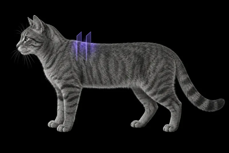
Why imaging comes before the first surgery
Feline injection-site sarcomas (FISS) develop at sites of previous vaccination or injection. According to the ABCD guidelines on FISS (Hartmann et al., Journal of Feline Medicine and Surgery, 2015), they behave more aggressively than fibrosarcomas elsewhere, and the rate of metastasis ranges from 10% to 28%. The guidelines state that:
- aggressive, radical excision is required to avoid recurrence
- prognosis improves when radiotherapy and/or immunotherapy are added
- injections should be given at sites where surgery would likely cure, generally avoiding the interscapular region
The first surgery matters. In 91 cats treated by radical excision with 5 cm margins and no adjunctive treatment (Phelps et al., Journal of the American Veterinary Medical Association, 2011), 13 (14%) had local recurrence and 18 (20%) developed metastasis. Median survival was 499 days with recurrence and 1,461 days without. In 22 cats treated with surgery and brachytherapy (Bloch et al., Journal of Feline Medicine and Surgery), time to progression was shorter after multiple surgeries than after a single surgery (310 days after multiple surgeries; no median could be given after a single surgery).
Imaging before the first excision is therefore worth discussing, so that the margins can be planned on the imaged extent of the tumour.
What CT shows
| Feature | Evidence |
|---|---|
| Location | 45% of 22 FISS fibrosarcomas were interscapular (Travetti et al., Journal of Feline Medicine and Surgery, 2013) |
| Enhancement | All but one enhanced strongly: ring (42%), heterogeneous (36%), homogeneous (9%) or mixed patterns (Travetti et al.) |
| Shape | Irregular in 62.5% of 32 cats, with digitiform projections in all (Zardo et al., Journal of Feline Medicine and Surgery) |
| Surrounding tissue | Blurred fat planes (68.8%) and liquefactive necrosis (68.8%) (Zardo et al.); marked invasion of musculature (Travetti et al.) |
| Skip metastases | Highly correlated with recurrence (P = 0.001) (Zardo et al.) |
In the fibrosarcoma study, the median CT volume was 7.57 cm³, and the longest axis ran craniocaudally in 68%.
CT measures more than the hand
In 53 cats with newly diagnosed FISS (Ferrari et al., Veterinary and Comparative Oncology), CT measurements of length and width tended to be larger than clinical measurements, and the difference grew with tumour size. Lateral and deep margins depend on these dimensions, so planning from palpation alone may underestimate the extent, especially in large tumours. The authors recommend that studies state which method defined tumour size.
Limits: peritumoral changes and grade
CT finds more than tumour. In 10 cats imaged with dual-phase CT angiography (CTA) and MRI before wide excision (Nemanic et al., Veterinary Surgery, 2016), 87 peritumoral lesions were examined microscopically:
- 17 (20%) were neoplastic
- 51 (59%) were non-neoplastic
- 19 (22%) were equivocal
CTA and MRI found a similar number of lesions. Imaging features overlapped so much that neoplastic peritumoral lesions could not be identified with certainty on either modality.
A secondary analysis of 10 cats from an earlier prospective study (Fleming et al., Veterinary Radiology & Ultrasound, 2019) found at least one neoplastic peritumoral projection in 6 cats on MRI, 5 on CTA and 3 on both. Imaging features did not predict tumour type or grade. On low-field MRI (Rousset et al., Veterinary Radiology & Ultrasound, 2013), the appearance was affected by previous incisional or excisional biopsy, and tumour volume did not predict tumour-free margins.
Interscapular CT protocol at a glance
The studies here used contrast-enhanced CT, including dual-phase CT angiography. For interscapular tumours, an extra post-contrast scan in a second forelimb position helped show how the tumour relates to the scapula and muscles, in the fibrosarcoma study. CTA is recommended for cats with known metallic objects in the field, because metal made one MRI non-diagnostic in the CTA and MRI study.
What the radiologist looks at
- Extent in every direction. Length, width and depth, because CT tends to measure more than palpation.
- Muscle and scapula. Which muscles are invaded, since marked invasion of musculature is common.
- Projections and fat planes. Digitiform projections and blurred fat planes, which were frequent in FISS.
- Skip metastases. Separate foci away from the main mass, given their correlation with recurrence.
- Peritumoral changes. Described with caution, because many were not tumour on histology.
- The lungs. When the thorax is included, for lung metastasis.
What your report will include
Every Veheri case is read by a radiologist or neurologist, and when you order you can choose Diplomate-only reporting or the full specialist panel. You can chat directly with the specialist assigned to your case, and a simplified pet owner report helps you explain the findings to the owner.
In practice, CT maps the tumour and its relationship to muscle and bone. It does not give a grade or confirm whether a peritumoral change is tumour or inflammation. When you take the report forward:
- Surgical planning. When imaging in two forelimb positions showed fewer infiltrated structures, a less invasive approach to the underlying muscles and scapula was performed.
- Grade and margins come from histology. Imaging did not predict tumour type or grade, and tumour volume did not predict tumour-free margins.
- Stage the thorax. After surgery and brachytherapy, distant failure occurred in 13.6% of cats, due to lung metastasis. With metastasis reported in 10–28% of FISS, it is worth discussing whether the thorax should be included in the same CT study.
- Talking to the owner: the first surgery matters, because recurrence was linked to much shorter survival.
What to include when you request a CT report
- signalment: age, breed and sex
- injection and vaccination history at the site
- previous biopsies or surgeries, their type (incisional or excisional) and dates
- palpated tumour dimensions
- metallic objects, such as a microchip, near the mass
- whether the plan is surgery, radiotherapy or both
- whether the thorax should be staged in the same study
How Veheri can help
Veheri’s radiologists report veterinary CT studies remotely for clinics, and can support your team with CT scanning protocols and positioning for oncology CT. For related reading, see CT of the feline lungs and oncology imaging for staging and planning.
Sources
Each claim in this guide links to its source. The full citations:
- Hartmann K, Day MJ, Thiry E, et al. Feline injection-site sarcoma: ABCD guidelines on prevention and management. J Feline Med Surg. 2015;17(7):606-13. doi:10.1177/1098612X15588451
- Phelps HA, Kuntz CA, Milner RJ, et al. Radical excision with five-centimeter margins for treatment of feline injection-site sarcomas: 91 cases (1998-2002). J Am Vet Med Assoc. 2011;239(1):97-106. doi:10.2460/javma.239.1.97
- Bloch J, Rogers K, Walker M, et al. Treatment of feline injection-site sarcoma with surgery and iridium-192 brachytherapy: retrospective evaluation of 22 cats. J Feline Med Surg. 22(4):313-321 (published online 2019). doi:10.1177/1098612X19844345
- Travetti O, di Giancamillo M, Stefanello D, et al. Computed tomography characteristics of fibrosarcoma: a histological subtype of feline injection-site sarcoma. J Feline Med Surg. 2013;15(6):488-93. doi:10.1177/1098612X12472174
- Zardo KM, Damiani LP, Matera JM, Fonseca-Pinto AC. Recurrent and non-recurrent feline injection-site sarcoma: computed tomographic and ultrasonographic findings. J Feline Med Surg. 18(10):773-82 (published online 2015). doi:10.1177/1098612X15596200
- Ferrari R, Di Giancamillo M, Stefanello D, et al. Clinical and computed tomography tumour dimension assessments for planning wide excision of injection site sarcomas in cats: how strong is the agreement? Vet Comp Oncol. 15(2):374-382 (published online 2015). doi:10.1111/vco.12173
- Nemanic S, Milovancev M, Terry JL, et al. Microscopic evaluation of peritumoral lesions of feline injection site sarcomas identified by magnetic resonance imaging and computed tomography. Vet Surg. 2016;45(3):392-401. doi:10.1111/vsu.12448
- Fleming K, Nemanic S, Löhr CV, et al. CT angiography and MRI imaging features do not predict the tumor type and grade of feline injection site sarcoma. Vet Radiol Ultrasound. 2019;60(6):668-679. doi:10.1111/vru.12807
- Rousset N, Holmes MA, Caine A, et al. Clinical and low-field MRI characteristics of injection site sarcoma in 19 cats. Vet Radiol Ultrasound. 2013;54(6):623-9. doi:10.1111/vru.12067
Related reading
More imaging guides
All imaging guides
Thoracic CT in cats for lung tumours, airway and interstitial disease
What thoracic CT adds over radiographs in cats with lung tumours, bronchial or interstitial disease, how anaesthesia affects the lungs, and where CT stops.
CT of the feline mediastinum for a cranial mediastinal mass
What CT adds for a cat with a cranial mediastinal mass, how far it separates thymoma, lymphoma and cysts, and why cytology or histology still decides.
CT of the feline middle ear
When to image the feline tympanic bulla, how CT compares with radiography, ultrasound and MRI, and what CT shows in otitis media, polyps and middle ear neoplasia.