
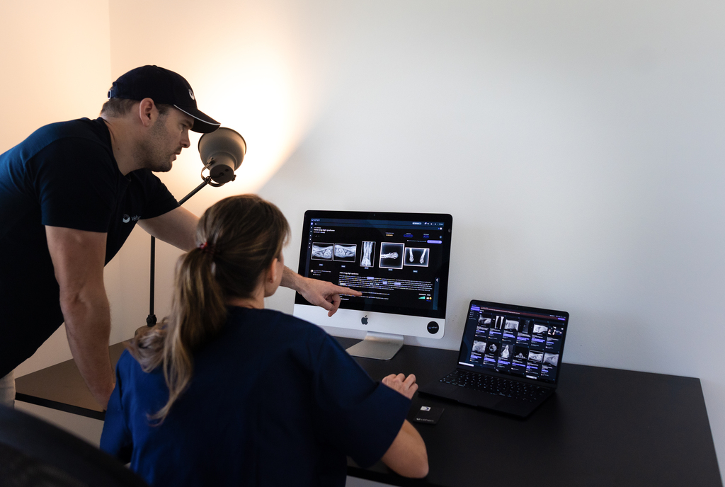
Veterinary radiology covers X-ray, ultrasound, CT, MRI, and nuclear medicine, interpreted by trained specialists (ACVR/ECVDI).
Follow ALARA with PPE, positioning aids, and sedation; standardize protocols and QC.
Use teleradiology for specialist reads and advanced cases, and refer for CT/MRI or complex differentials.
See ACVR, Merck Veterinary Manual, UC Davis CEH, AVMA, and ECVDI for authoritative guidance.
Veterinary Radiology: Modalities, Training, Safety, and When to Refer If you’re asking “what is veterinary radiology,” it’s the specialty that uses medical imaging (X-ray, ultrasound, CT, MRI, nuclear medicine) to diagnose disease in animals and guide treatment decisions.
Veterinary radiology spans everyday in-clinic radiographs and ultrasounds through advanced CT/MRI, plus teleradiology for specialist interpretations. Below, I explain the modalities, safety standards, training/accreditation, and how to decide when to refer.
What is veterinary radiology?
Veterinary radiology (often called veterinary diagnostic imaging) is a board-certified specialty that interprets medical images to diagnose disease in animals.
You can become a diplomate through advanced training in an accredited residency program, followed by board certification through organizations such as the American College of Veterinary Radiology (ACVR) or the European College of Veterinary Diagnostic Imaging (ECVDI), supported by professional associations like the EAVDI.
Core imaging modalities in veterinary practice
A quick orientation to the most common modalities, what they show best, and typical use-cases. Selecting the right test is about matching the question to each modality’s strengths.
- Radiography (X-ray)
First-line for bones, lungs, abdomen overview; fast, widely available. See modality basics, technique, positioning, and interpretation in the Merck Veterinary Manual.
- Ultrasound
Real-time soft-tissue evaluation of abdominal organs and superficial structures; with cardiac probes (echo) for heart structure and function.
- Computed Tomography (CT)
High-detail cross-sectional imaging of skull, thorax, abdomen, and extremities; excellent for complex fractures, nasal disease, and neoplasia staging; usually requires general anesthesia.
- Magnetic Resonance Imaging (MRI)
Gold standard for neurologic and many musculoskeletal soft-tissue lesions; superior soft-tissue contrast; anesthesia typically required.
- Nuclear medicine (scintigraphy) and PET
Functional imaging for bone turnover or metabolic activity; used more commonly in equine lameness and oncology centers. For pros/cons and use-cases, see UC Davis CEH Imaging.
“Radiology:Veterinary specialists who focus on the study of X-ray, ultrasound, computed tomography (CT), magnetic resonance imaging (MRI), and other imaging procedures that allow us to see “inside” an animal’s body; radiation oncologists specialize in treating cancer patients using radiotherapy.— AVMA: What board-certified veterinary specialists do
Cross-sectional vs. functional imaging
CT and MRI are cross-sectional modalities that reveal anatomy with high detail; scintigraphy and PET demonstrate physiology (e.g., bone turnover, metabolic activity), so they answer different clinical questions and often complement each other.
ALSO READ: How to find an appropriate veterinary CT Scanner
Safety and the ALARA principle
Radiation safety underpins responsible imaging. Following ACVR guidance, practices should use proper PPE, collimation, exposure protocols, and routine equipment QC. Sedation and positioning aids reduce manual restraint and exposure.
“Exposure to radiation should always be As Low As Reasonably Achievable (ALARA) while maximizing the quality of the procedure.” — ACVR: Radiation Safety Statement
“PPE including a lead (or equivalent) apron, thyroid collar, fully enclosing gloves (worn on the hands, not draped over them), and lead glasses should be worn at all times while remaining in the room with a patient for a procedure involving ionizing radiation.” — ACVR: Radiation Safety Statement
For technique, positioning, contrast use, digital workflows (DICOM/PACS), and interpretation, see Merck Veterinary Manual.
ALARA in practice
Use a technique chart, strict collimation, sedation to minimize manual restraint, and yearly PPE checks. Monitor staff exposure with dosimetry according to local regulations and ACVR recommendations.
Training and certification: ACVR and ECVDI
Specialists train via residencies and board exams. In North America, ACVR board-certifies in Diagnostic Imaging and Equine Diagnostic Imaging. In Europe, ECVDI (recognized by EBVS) certifies small- and large-animal tracks with theoretical and practical exams.
Learn more: ACVR: Types of Imaging & Therapy and ACVR: Diplomate Accreditation. European perspective: ECVDI Overview and ECVDI Resident Exams.
Pathways: ACVR vs ECVDI
Both require supervised residency training, case logs, scholarly activity, and passing rigorous board examinations. ACVR offers Diagnostic Imaging and Equine DI; ECVDI offers small- and large-animal tracks with January/February exam sessions.
When to refer, and how teleradiology helps
General practices can handle many X-rays and ultrasounds in-house. Refer or seek a specialist read when:
- The study is non-diagnostic yet repeating will add risk/cost.
- Advanced imaging (CT/MRI) is indicated for surgical planning, neurologic disease, staging, or complex lameness.
- You need high-stakes, nuanced differentials or treatment-impacting details.
According to the Merck Veterinary Manual, the internet enables sending images to a radiologist for interpretation. Teleradiology lets clinics upload DICOM studies securely for board-certified interpretation, accelerating diagnosis and care while minimizing repeat exposures.
Smarter Teleradiology. Faster Diagnosis. Better Results Join the Veheri Community! Start today: Book your DEMO
Imaging guides on this topic
Browse all imaging guides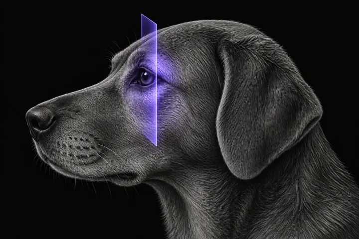
CT of the canine orbit for exophthalmos and retrobulbar disease
How CT helps separate orbital neoplasia from cellulitis and other inflammation in dogs with exophthalmos, where MRI adds detail, and how CT guides sampling.
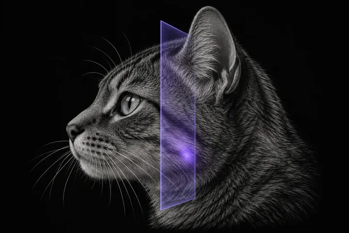
CT of the feline middle ear
When to image the feline tympanic bulla, how CT compares with radiography, ultrasound and MRI, and what CT shows in otitis media, polyps and middle ear neoplasia.
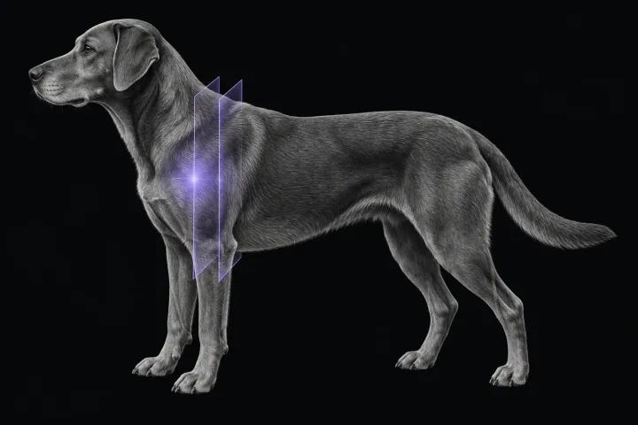
MRI of the canine shoulder for biceps and supraspinatus tendinopathy
What MRI shows in canine biceps and supraspinatus tendinopathy, how it compares with CT arthrography, and why a bright tendon alone is not disease.