
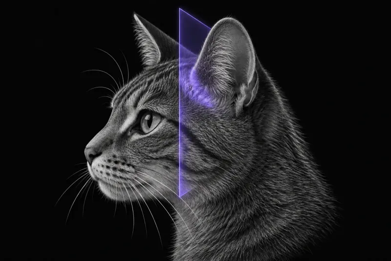
Start with the localisation
The first imaging question in a vestibular cat is whether the lesion is central or peripheral, because the likely causes differ.
In 77 cats with vestibular signs that had MRI of the head (Negrin et al., Journal of Feline Medicine and Surgery, 2010), 40 (52%) had central vestibular dysfunction. Its most frequent causes were:
- inflammatory disease (18 cats), including intracranial extension of otitis interna (5), feline infectious peritonitis (3) and toxoplasmosis (2)
- neoplasia (12)
- vascular disease (4)
Of 37 cats with peripheral dysfunction, idiopathic vestibular syndrome was suspected in 16 and otitis media/interna in 16. Where MRI showed a lesion, its location agreed with the clinical localisation in 52 of 55 cats (95%). Localisation was a significant predictor of survival.
What peripheral vestibular syndrome turns out to be
In 196 cats with peripheral vestibular syndrome, all imaged with CT or MRI (Caldero Carrete et al., Veterinary Record, 2025), the diagnoses were:
| Diagnosis | Cats |
|---|---|
| Otitis media/interna alone | 91 |
| Otitis media/interna with aural polyps | 49 |
| Idiopathic vestibular syndrome | 47 |
| Middle ear neoplasia | 7 |
| Congenital vestibular syndrome | 2 |
Otitis media/interna was associated with younger age, a longer duration of signs, a history of otitis externa or upper respiratory signs, facial nerve paralysis and Horner syndrome.
MRI compared with CT
The comparison below follows an image essay on otitis media in dogs and cats (Ludewig et al., Tierärztliche Praxis Ausgabe K: Kleintiere / Heimtiere, 2024).
| Question | Better answered by |
|---|---|
| Bulla wall changes and abnormal bulla content | CT is more sensitive |
| Guiding sampling of the bulla | CT is often easier |
| Nature of the material inside the bulla | MRI |
| Meninges, brain and cranial nerves; central spread of otitis media | MRI is clearly superior |
In practice, CT answers the bony middle ear question, while MRI is usually preferred when signs are central, when intracranial extension is suspected, or when the bulla looks normal and the inner ear itself is the question.
Idiopathic vestibular syndrome
In a survey of neurology specialists (Mertens et al., Frontiers in Veterinary Science, 2023), idiopathic vestibular syndrome was defined as an acute to peracute, improving, non-painful peripheral vestibular disorder that affects cats of any age. A full work-up may include MRI and CSF analysis to rule out other causes. Specialists in the EU used advanced imaging less often and otoscopy more often.
Inner ear MRI protocol at a glance
FLAIR images are central to inner ear MRI, because normal perilymph is expected to be suppressed on FLAIR (Everest et al., Journal of Feline Medicine and Surgery, 2023). Post-contrast images show meningeal enhancement, and the T1-weighted sequence matters: observers detected it more often on fat-suppressed turbo spin echo than on 3D gradient echo images (Graf et al., Frontiers in Veterinary Science, 2025). When the bulla needs sampling, CT is often easier.
What the radiologist looks at
- Where the lesion is. Central or peripheral, which drives the list of likely causes.
- FLAIR suppression. Perilymph is a very low-protein fluid. In 41 cats, the inner ear signal was significantly less suppressed in cats with clinical otitis media/interna than in cats with inflammatory brain disease, non-inflammatory structural disease or a normal brain MRI.
- Idiopathic vestibular syndrome is different. In 25 cats with idiopathic vestibular syndrome and clearly lateralised signs (Putzer et al., Journal of Feline Medicine and Surgery, 2026), the side with reduced FLAIR suppression matched the clinical side in 14 and did not in 11. There was no correlation, unlike a previous report in dogs.
- Meningeal enhancement. In 58 cats with otitis media/interna that had post-contrast MRI and CSF analysis (Dutil et al., Journal of Feline Medicine and Surgery, 2022), meningeal enhancement was reported in 26. It was not associated with CSF abnormalities or bacteriology results, and outcome tended to be poorer when it was present.
- Over-reading. In 74 cats and 46 dogs with otitis media/interna, the difference between sequences was significant in cats only, and the authors warn that normal meninges can be over-read.
What your report will include
Every Veheri case is read by a radiologist or neurologist, and when you order you can choose Diplomate-only reporting or the full specialist panel. Neurology reporting covers CT and MRI. You can chat directly with the specialist assigned to your case, and a simplified pet owner report helps you explain the findings to the owner.
When you take the report forward:
- The affected side in idiopathic vestibular syndrome cannot be shown reliably on MRI.
- Meningeal enhancement does not predict CSF results. CSF abnormalities were more common in acute (16 of 37) than chronic cases (3 of 21).
- The FLAIR findings come from retrospective studies of presumptive diagnoses: the 41-cat study and the 25-cat study.
- Talking to the owner: of 104 cats with peripheral vestibular syndrome and follow-up, 33 recovered fully, 67 partially and 4 not at all. In the 77-cat MRI series, 9 of 16 cats with presumed idiopathic disease (56%) recovered rapidly and completely. Most of these cats had signs that progressed over 3 weeks and were classed as atypical.
What to include when you request an MRI report
- signalment: age, breed and sex
- head tilt direction, nystagmus type and direction, mentation, postural reactions and other cranial nerve deficits, to support central or peripheral localisation
- facial nerve paralysis or Horner syndrome
- history of otitis externa or upper respiratory signs, and otoscopic findings
- onset, duration and progression of signs
- CSF results if available
How Veheri can help
Veheri works with veterinary radiologists and neurologists and reports head MRI and CT studies remotely for clinics, with expert support for scanning protocols. For the bony middle ear, see CT of the feline middle ear, and for dogs, MRI of the inner ear and vestibular system in dogs.
Sources
Each claim in this guide links to its source. The full citations:
- Negrin A, Cherubini GB, Lamb C, et al. Clinical signs, magnetic resonance imaging findings and outcome in 77 cats with vestibular disease: a retrospective study. J Feline Med Surg. 2010;12(4):291-9. doi:10.1016/j.jfms.2009.10.001
- Caldero Carrete J, De Decker S, Volk HA, et al. Peripheral vestibular syndrome in cats: clinical presentation, diagnostic findings and outcome in 196 cases. Vet Rec. 2025;197(9):e5324. doi:10.1002/vetr.5324
- Ludewig E, Jopp I, Vali Y. Imaging findings in otitis media and resulting secondary lesions in dogs and cats - an image essay. Tierarztl Prax Ausg K Kleintiere Heimtiere. 2024;52(3):151-161. doi:10.1055/a-2324-0318
- Everest S, Monteith G, Gaitero L, Samarani F. Suppression of inner ear signal intensity on fluid-attenuated inversion recovery magnetic resonance imaging in cats with vestibular disease. J Feline Med Surg. 2023;25(4):1098612X231168001. doi:10.1177/1098612X231168001
- Putzer M, De Decker S, Gutierrez Quintana R, et al. Lack of association between inner ear FLAIR suppression on MRI and laterality of clinical signs in feline idiopathic vestibular syndrome. J Feline Med Surg. 2026;28(7):1098612X261445137. doi:10.1177/1098612X261445137
- Dutil GF, Guevar J, Schweizer D, et al. Otitis media and interna with or without polyps in cats: association between meningeal enhancement on postcontrast MRI, cerebrospinal fluid abnormalities, and clinician treatment choice and outcome. J Feline Med Surg. 2022;24(12):e481-e489. doi:10.1177/1098612X221125573
- Graf F, Dennler M, Beckmann K, et al. Comparative study of meningeal enhancement in canine and feline otitis media-interna: 3D-gradient-echo vs. fat-suppressed turbo-spin-echo-T1-weighted sequences in MRI. Front Vet Sci. 2025;12:1664006. doi:10.3389/fvets.2025.1664006
- Mertens AM, Schenk HC, Volk HA. Current definition, diagnosis, and treatment of canine and feline idiopathic vestibular syndrome. Front Vet Sci. 2023;10:1263976. doi:10.3389/fvets.2023.1263976
Related reading
More imaging guides
All imaging guides
MRI of the inner ear and vestibular system in dogs
How reliably the neurological exam separates central from peripheral vestibular disease in dogs, and what MRI shows in otitis interna and idiopathic cases.
Brain MRI in cats
What brain MRI shows in cats with seizures, meningioma, lymphoma or FIP, how often the scan is normal, and what MRI cannot settle on its own.
Pituitary imaging for hypersomatotropism in cats
When diabetic cats need pituitary imaging for hypersomatotropism, what MRI and CT show, and why a normal-sized gland does not rule it out.