
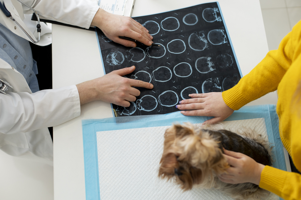
Expert vet neuroimaging reports by specialists—fast, reliable, and actionable.
Expert vet neuroimaging reports by specialists—fast, reliable, and actionable. Discover the Veheri difference in vet neuroimaging: board-certified neurologists, rapid turnaround, personalized therapy recommendations, and seamless collaboration for optimal veterinary care.
Veheri Neuroimaging Reports: Setting a New Standard in Vet Neuroimaging In the fast-evolving world of veterinary medicine, the demand for advanced diagnostic tools and expert interpretation has never been higher. When it comes to Vet Neuroimaging, accuracy, speed, and clinical insight are essential for delivering the best possible care to animal patients. At Veheri, we are proud to offer a neuroimaging reporting service that stands out for its quality, efficiency, and support for veterinarians.
Unmatched Expertise: Reports by Board-Certified Veterinary Neurologists
At Veheri, all neurology reports are written exclusively by board-certified veterinary neurologists (Diplomates), ensuring the highest professional and diagnostic standards.
Our commitment to excellence begins with our team. Every report is crafted by a board-certified veterinary neurologist, bringing years of specialized training and clinical experience to each case. This ensures that every interpretation is not only precise but also grounded in the latest advances in Vet Neuroimaging and neurology.
Rapid Turnaround: When Every Minute Counts
We understand that time is often of the essence in veterinary practice. That’s why our standard reports are typically completed within one working day, allowing you to move forward with your cases without unnecessary delays. For urgent situations, our express option delivers results in an average of just six hours—helping you make critical decisions faster and with confidence.
Beyond the Basics: Personalized Therapy Recommendations
At Veheri, we believe that a neuroimaging report should be more than just a list of findings. Each report includes a personalized therapy recommendation, tailored to the specific needs of your patient. This added layer of clinical insight supports your next steps, whether it’s further diagnostics, medical management, or surgical intervention.
Comprehensive Service for Better Veterinary Care
Our comprehensive approach to Vet Neuroimaging empowers veterinarians to manage cases rapidly and make evidence-based therapy decisions. By outsourcing image interpretation to our team of specialists, you free up valuable time to focus on what matters most—caring for your patients. Meanwhile, you can trust that you’re receiving a precise, reliable diagnosis that meets the highest standards of veterinary care.
Seamless Collaboration: Direct Access to Neurologists
Communication is key to optimal patient outcomes. That’s why Veheri offers an integrated chat function, allowing you to connect directly with the neurologist responsible for your case. Whether you have follow-up questions or need clarification on a report, our team is just a message away, ensuring smooth collaboration and peace of mind.
Why Veheri is the Smart Choice for Vet Neuroimaging
- Reports exclusively by board-certified veterinary neurologists
- Fast, reliable turnaround—standard or express
- Personalized, actionable therapy recommendations
- Direct communication with your reporting neurologist
- More time for you to focus on patient care
- Confidence in every diagnosis
Experience the Veheri Difference. At Veheri, we are dedicated to supporting veterinarians with the highest quality neuroimaging reports and expert guidance. Our mission is to make advanced Vet Neuroimaging accessible, efficient, and clinically valuable—so you can deliver the best possible care to your patients.
Smarter Teleradiology. Faster Diagnosis. Better Results
Join the Veheri Community!
Start today: Book your DEMO
Imaging guides on this topic
Browse all imaging guides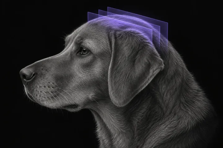
Brain MRI for dogs with seizures
Which dogs with seizures need brain MRI, what the evidence says about diagnostic yield, what the radiologist looks for and what your report will include.
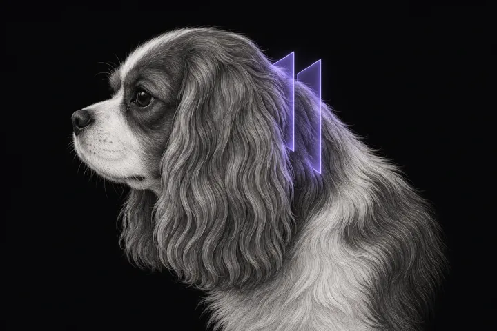
MRI of the craniocervical junction for Chiari-like malformation and syringomyelia
What MRI of the canine craniocervical junction should include for Chiari-like malformation and syringomyelia, and how to read a syrinx against the signs.
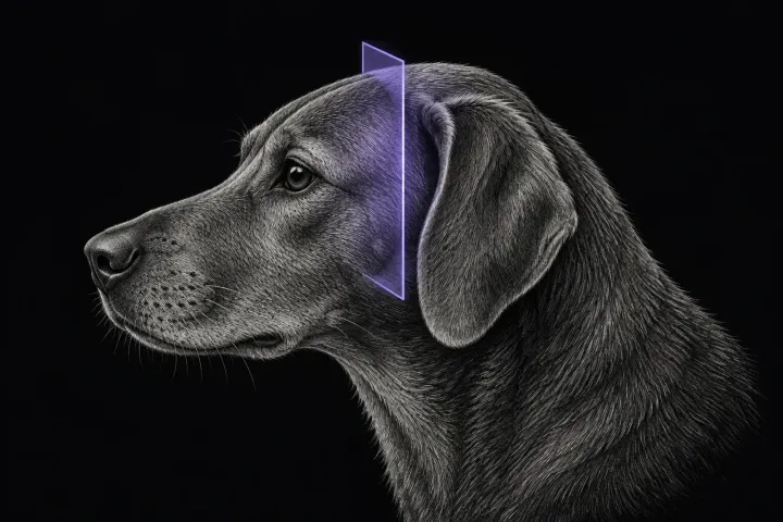
MRI of the trigeminal nerve in dogs with a dropped jaw or masticatory atrophy
How MRI separates idiopathic trigeminal neuropathy, neuritis and nerve sheath tumours in dogs with a dropped jaw or masticatory atrophy, and its pitfalls.