
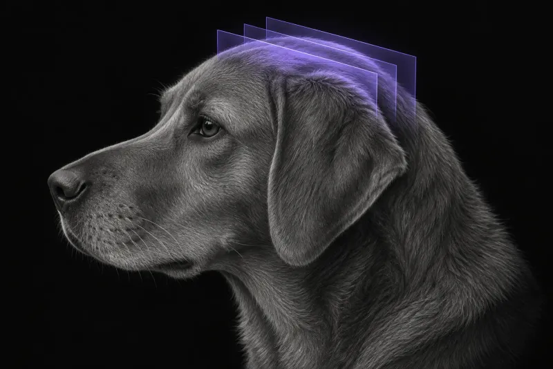
What MRI is for in a seizure patient
For a dog with epileptic seizures, brain MRI has three aims, as the International Veterinary Epilepsy Task Force (IVETF) sets out in its MRI protocol recommendations (Rusbridge et al., BMC Veterinary Research, 2015):
- find causes that need more than antiepileptic treatment, such as inflammatory or infectious brain disease
- identify lesions that are a consequence of seizures rather than their cause
- support research into epilepsy
Idiopathic epilepsy is a diagnosis of exclusion, so the MRI of a dog with idiopathic epilepsy is expected to be normal. MRI must always follow a good history and a clinical and neurological examination, and a normal MRI does not predict prognosis or tell you which drug will work.
Which dogs need MRI
The IVETF consensus on diagnosing epilepsy in dogs (De Risio et al., BMC Veterinary Research, 2015) uses three tiers of confidence for an idiopathic epilepsy diagnosis:
| Tier | Based on |
|---|---|
| I | Two or more unprovoked epileptic seizures at least 24 h apart; onset between 6 months and 6 years of age; unremarkable interictal physical and neurological examination; no significant abnormalities on minimum database blood tests and urinalysis |
| II | Tier I, plus unremarkable fasting and post-prandial bile acids, brain MRI using an epilepsy-specific protocol, and CSF analysis |
| III | Tiers I and II, plus EEG abnormalities characteristic of seizure disorders |
After reactive seizures have been excluded, the IVETF recommends brain MRI and routine CSF analysis when a dog has any of these red flags:
- seizure onset before 6 months or after 6 years of age
- interictal neurological abnormalities consistent with an intracranial lesion
- status epilepticus or cluster seizures at onset
- a previous presumptive diagnosis of idiopathic epilepsy, with drug resistance to a single antiepileptic drug titrated to the highest tolerable dose
flowchart TD
  accTitle: Does this dog with seizures need brain MRI?
  accDescr: Decision path from the IVETF consensus, once reactive seizures are ruled out. Any red flag leads to brain MRI and CSF analysis. Without one, idiopathic epilepsy can be diagnosed at Tier I confidence, and brachycephalic dogs deserve a lower threshold for MRI.
  start["Seizures, reactive causes ruled out"] --> flags{{"Any IVETF red flag?"}}
  flags -- "Yes" --> mri["Brain MRI + CSF analysis"]
  flags -- "No" --> tier1["Tier I idiopathic epilepsy if criteria met"]
tier1 -. "Brachycephalic" .-> mriFor other neurological presentations, see when veterinary MRI is the right move.
What the evidence says about diagnostic yield
- Age and the neurological exam. A study of epileptic dogs without interictal deficits (Smith et al., The Veterinary Journal) found significant MRI abnormalities in 1 of 46 dogs younger than 6 years (2.2%) but in 8 of 30 dogs older than 6 years (26.7%).
- Seizure pattern. In 404 dogs with seizures of intracranial origin (Armaşu et al., The Veterinary Journal, 2014), older age at onset made an asymmetrical structural lesion significantly more likely. Dogs with single seizures were less likely to have one than dogs with cluster seizures.
- Skull shape. In a 2025 single-centre study of 111 dogs with generalised seizures (Prodger et al., Journal of Small Animal Practice), a structural cause was found in 61.8% of brachycephalic dogs versus 22.1% of other dogs. Among dogs aged 6 months to 6 years with a normal interictal examination, 33.3% of brachycephalic dogs had a structural lesion, against none of the other dogs. The authors suggest MRI and CSF analysis should be more strongly considered in brachycephalic dogs that otherwise meet Tier I criteria.
Brain MRI protocol at a glance
Brain protocols still differ between institutions, but most share the same core. T2-weighted and T1-weighted sagittal sequences are run on both high-field and low-field scanners. FLAIR and gradient echo sequences are common additions, diffusion-weighted imaging is useful on a high-field system, and post-contrast sequences are recommended when there is doubt. For seizure patients, the IVETF has published an epilepsy-specific protocol built around the hippocampus.
What the radiologist looks at
The study is read against the history you send, because the same signal change can mean different things in different patients. The points below follow the IVETF protocol recommendations.
- Post-ictal change or a lesion? Seizure-induced changes most often appear as T2-weighted hyperintensities in the piriform and temporal lobes, the cingulate gyrus and the hippocampus, sometimes with mild contrast uptake. Their diffuse pattern and typical location usually separate them from an epileptogenic lesion.
- The hippocampus. At least a visual assessment for atrophy, asymmetry, loss of structure, increased T2-weighted or FLAIR signal and decreased T1-weighted signal.
- Is a lesion the cause? A lesion is not automatically epileptogenic. Other evidence, such as EEG or the seizure history, may be needed to link it to the seizures.
What your report will include
Every Veheri case is read by a radiologist or neurologist, and when you order you can choose Diplomate-only reporting or the full specialist panel. Neurology reporting covers CT and MRI, and neurology reports include a personalised therapy recommendation. You can chat directly with the specialist assigned to your case, and a simplified pet owner report helps you explain the findings to the owner.
When you take the report forward:
- A normal scan fits idiopathic epilepsy, which is a diagnosis of exclusion. It supports a Tier II diagnosis when the bile acids and CSF analysis are also unremarkable.
- Suspected post-ictal change may need a repeat scan later, because these changes usually resolve within 16 weeks.
- CSF taken soon after a seizure can mislead, because post-ictal pleocytosis can occur.
- Talking to the owner: a normal MRI does not predict prognosis or which drug will work, so set expectations about treatment response separately.
What to include when you request a brain MRI report
- signalment: age, breed and sex
- age at seizure onset, seizure type, frequency, and any clusters or status epilepticus
- interictal neurological examination findings
- minimum database, bile acids and CSF results where available
- current antiepileptic drugs, doses and response
- time since the last seizure, so post-ictal changes can be interpreted
How Veheri can help
Veheri works with veterinary radiologists and neurologists and reports brain MRI studies remotely for clinics. For cats, see brain MRI in cats, and for clinics without MRI, brain CT in dogs when MRI is not available.
Sources
Each claim in this guide links to its source. The full citations:
- De Risio L, Bhatti S, Muñana K, et al. International Veterinary Epilepsy Task Force consensus proposal: diagnostic approach to epilepsy in dogs. BMC Vet Res. 2015;11:148. doi:10.1186/s12917-015-0462-1
- Rusbridge C, Long S, Jovanovik J, et al. International Veterinary Epilepsy Task Force recommendations for a veterinary epilepsy-specific MRI protocol. BMC Vet Res. 2015;11:194. doi:10.1186/s12917-015-0466-x
- Smith PM, Talbot CE, Jeffery ND. Findings on low-field cranial MR images in epileptic dogs that lack interictal neurological deficits. Vet J. 176(3):320-5 (published online 2007). doi:10.1016/j.tvjl.2007.03.003
- Armaşu M, Packer RM, Cook S, et al. An exploratory study using a statistical approach as a platform for clinical reasoning in canine epilepsy. Vet J. 2014;202(2):292-6. doi:10.1016/j.tvjl.2014.08.008
- Prodger A, Khan S, Harris G. Prevalence of structural and idiopathic epilepsy in brachycephalic and non-brachycephalic dogs in the context of the International Veterinary Epilepsy Task Force guidelines. J Small Anim Pract. 2025;66(8):540-546. doi:10.1111/jsap.13857
Related reading
More imaging guides
All imaging guides
Brain CT in dogs when MRI is not available
What brain CT can and cannot detect in dogs when MRI is not available, for masses, stroke, inflammation and head trauma, and when MRI or CSF is still needed.
Brain MRI in cats
What brain MRI shows in cats with seizures, meningioma, lymphoma or FIP, how often the scan is normal, and what MRI cannot settle on its own.
MRI of the canine brachial plexus for peripheral nerve sheath tumours
How MRI finds peripheral nerve sheath tumours of the canine brachial plexus, what the radiologist looks for, and what imaging can say about grade and extent.