
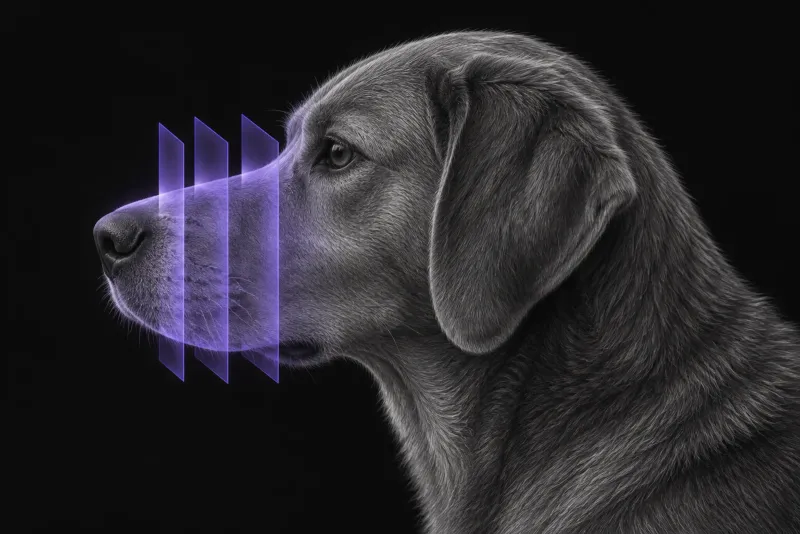
Why chronic nasal discharge deserves a work-up
Chronic nasal discharge is often treated first with antibiotics, yet it is frequently the sign of a serious primary disease. In a referral series of 85 dogs with nasal discharge (Rösch et al., Tierärztliche Praxis Ausgabe K: Kleintiere / Heimtiere, 2019), the underlying causes were:
- nasal neoplasia (28%)
- oronasal defects (26%)
- idiopathic chronic rhinitis (20%)
- foreign bodies (10%)
- sinonasal aspergillosis (8%)
- diseases of the nasal planum (8%)
No dog had a primary bacterial infection, although 72% had already received antibiotics. In brachycephalic dogs, oronasal defects were the most frequent cause (8 of 14).
The same authors conclude that antibiotics without further diagnostics are not appropriate, and can be harmful in dogs with nasal tumours or aspergillosis. They also found that culture of nasal swabs did not identify the primary disease.
When CT is indicated
CT is indicated when nasal signs persist or recur and a structural cause is possible:
- unilateral or bilateral discharge that does not resolve
- epistaxis
- facial deformity
- a suspected foreign body that is not visible
CT defines the extent of disease in the palate, nasopharyngeal meatus, maxillary sinus, caudal ethmoturbinates and periorbital tissues. These areas are difficult to assess on radiographs (Codner et al., Journal of the American Veterinary Medical Association, 1993).
How accurate CT is
In a retrospective study of 80 dogs with chronic nasal disease (Saunders et al., Veterinary Radiology & Ultrasound, 2003), CT diagnosis had an accuracy above 90% for nasal neoplasia, nasal aspergillosis, non-specific rhinitis and foreign-body rhinitis. Agreement between two observers was substantial to almost perfect.
Nasal tumours: CT staging and prognosis
When a tumour is confirmed, CT is also the staging tool. In 94 dogs treated with intent-to-cure radiotherapy (Adams et al., Veterinary Radiology & Ultrasound, 2009), a modified CT-based staging system was prognostically significant:
- dogs with unilateral intranasal disease and no bone destruction beyond the turbinates had the longest median survival (23.4 months)
- CT evidence of cribriform plate involvement was associated with the shortest (6.7 months)
- combining CT stage with histology improved prognostic value
Nasal CT protocol at a glance
Nasal CT is usually done under general anaesthesia in the same episode as rhinoscopy and sampling, as in the referral series, an aspergillosis diagnosis study (Pomrantz et al., Journal of the American Veterinary Medical Association, 2007) and a study of blood ratios in nasal disease (Rösch et al., BMC Veterinary Research, 2024). This spares the patient a second anaesthetic and lets the rhinoscopist target the lesions CT has already mapped. The scan has to cover every area the report must describe, from the nasal cavity to the cribriform plate, orbit, frontal sinus, nasopharynx and regional lymph nodes.
What the radiologist looks at
The radiologist reads the morphology and distribution of the disease. An earlier comparison of CT with radiography described the two main patterns that separate tumours from infectious rhinitis:
| Finding on CT | Points towards |
|---|---|
| Space-occupying lesion that obliterates the turbinates, deviates the nasal septum and erodes bone | Nasal neoplasia |
| Cavitating lesion that spares the paranasal sinuses, with thickened, distorted turbinates and a widened meatus | Infectious rhinitis (bacterial or fungal in that series) |
Two limits matter in practice:
- Aspergillosis confined to the nasal passages can look like foreign-body rhinitis when the foreign body itself is not visible.
- Attenuation values and the degree of contrast enhancement did not separate infectious rhinitis from neoplasia in the radiography comparison.
When a tumour is likely, the report should describe explicitly the integrity of the cribriform plate, extension into the orbit, frontal sinus or nasopharynx, and the regional lymph nodes, because cribriform plate involvement carried the shortest survival.
What your report will include
Every Veheri case is read by a radiologist or neurologist, and when you order you can choose Diplomate-only reporting or the full specialist panel. You can chat directly with the specialist assigned to your case, and a simplified pet owner report helps you explain the findings to the owner.
CT narrows the differential and shows where to sample. It does not replace tissue diagnosis. When you take the report forward:
- Biopsy a suspected tumour under endoscopic guidance. When a malignant intranasal tumour is suspected, endoscopic-guided biopsies of the mass are recommended, because blind nasal biopsies carry a high false-negative rate.
- Confirm aspergillosis with more than one test. In the prospective serology study, serum anti-Aspergillus antibodies had a sensitivity of 67% and specificity of 98%, and fungal culture of nasal tissue a sensitivity of 81% and specificity of 100%. A positive result strongly supports the diagnosis; a negative result does not rule it out. That study defined aspergillosis by at least two of: consistent CT findings, fungal plaques on rhinoscopy and fungal hyphae on histology.
- Treat blood tests as a clue only. Routine haematology is usually only mildly abnormal in nasal disease. A raised platelet-to-lymphocyte ratio was associated with malignant disease and may serve as an additional screening clue, not a diagnosis.
- Talking to the owner: it can help to explain why antibiotics alone are not enough. The referral series authors consider antibiotics without further diagnostics inappropriate, and potentially harmful with tumours or aspergillosis.
What to include when you request a nasal CT report
- signalment: age, breed and sex
- duration and laterality of the discharge, and whether it is serous, mucopurulent or haemorrhagic
- epistaxis, sneezing, stertor, facial swelling or pain on palpation of the nasal planum
- dental history, since oronasal defects are a common cause
- previous treatment and response, including antibiotics
- breed conformation, because brachycephalic dogs have a different mix of causes
How Veheri can help
Veheri provides veterinary teleradiology and reports head CT studies remotely for clinics. For cats, see CT of the feline nasal cavity, and for tumour staging more widely, our practical guide to oncology imaging.
Sources
Each claim in this guide links to its source. The full citations:
- Saunders JH, van Bree H, Gielen I, de Rooster H. Diagnostic value of computed tomography in dogs with chronic nasal disease. Vet Radiol Ultrasound. 2003;44(4):409-13. doi:10.1111/j.1740-8261.2003.tb00477.x
- Codner EC, Lurus AG, Miller JB, et al. Comparison of computed tomography with radiography as a noninvasive diagnostic technique for chronic nasal disease in dogs. J Am Vet Med Assoc. 1993;202(7):1106-10. PMID: 8473224. https://pubmed.ncbi.nlm.nih.gov/8473224/
- Rösch S, Bomhard WV, Heilmann RM, Oechtering GU. Nasal discharge in dogs – are microbiological and histopathological examinations clinically useful? Tierarztl Prax Ausg K Kleintiere Heimtiere. 2019;47(2):84-96. doi:10.1055/a-0863-6667
- Pomrantz JS, Johnson LR, Nelson RW, Wisner ER. Comparison of serologic evaluation via agar gel immunodiffusion and fungal culture of tissue for diagnosis of nasal aspergillosis in dogs. J Am Vet Med Assoc. 2007;230(9):1319-23. doi:10.2460/javma.230.9.1319
- Adams WM, Kleiter MM, Thrall DE, et al. Prognostic significance of tumor histology and computed tomographic staging for radiation treatment response of canine nasal tumors. Vet Radiol Ultrasound. 2009;50(3):330-5. doi:10.1111/j.1740-8261.2009.01545.x
- Rösch S, Woitas J, Neumann S, et al. Diagnostic benefits of platelet-to-lymphocyte, neutrophil-to-lymphocyte, and albumin-to-globulin ratios in dogs with nasal cavity diseases. BMC Vet Res. 2024;20(1):44. doi:10.1186/s12917-024-03876-5
Related reading
More imaging guides
All imaging guides
CT of the feline nasal cavity for chronic nasal disease
Which CT features separate nasal lymphoma, carcinoma, fungal and chronic rhinitis in cats, why the retropharyngeal nodes matter, and why biopsy is still needed.

CT of adrenal masses in dogs
What contrast CT shows in a dog with an adrenal mass, from caval invasion and adhesions to tumour type, and where its accuracy runs out before adrenalectomy.

Brain CT in dogs when MRI is not available
What brain CT can and cannot detect in dogs when MRI is not available, for masses, stroke, inflammation and head trauma, and when MRI or CSF is still needed.