
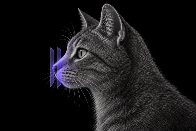
Why chronic nasal disease in cats needs imaging
Chronic rhinitis, nasal lymphoma, carcinoma and fungal disease can all cause the same signs in cats. In 167 cats seen for nasal or nasopharyngeal disease (Vilcot et al., Journal of Veterinary Internal Medicine, 2026), systemic signs were associated with nasal masses, while stertor and dyspnoea pointed to the nasopharynx. Clinical signs help to localise the disease, but they do not give a diagnosis.
CT shows where the disease is and how far it extends. In 62 cats with sinonasal disease (Schoenborn et al., Veterinary Radiology & Ultrasound, 2003), CT was not more sensitive than radiographs at detecting nasal abnormalities, but it was more sensitive at localising them and defining their extent.
CT features that point towards neoplasia
Three retrospective studies looked at CT features against histology. Together they describe a consistent pattern:
| CT feature | Association |
|---|---|
| Osteolysis of the paranasal bones, moderate to severe turbinate destruction, nasal septum lysis, a homogeneous mass, extension into the orbit or facial soft tissue | Findings that may lead to a CT diagnosis of neoplasia in the 62-cat study |
| Unilateral ethmoturbinate lysis; lysis of the vomer and of the ventral, dorsal and lateral maxilla | Neoplasia, in 43 cats, 15 with neoplasia and 28 with rhinitis (Tromblee et al., Veterinary Radiology & Ultrasound, 2006) |
| Unilateral soft tissue or fluid in the sphenoid sinus, frontal sinus or retrobulbar space | Neoplasia, in the same 43 cats |
| A nasal mass, turbinate lysis or paranasal bone lysis | Neoplasia, in 56 cats, 22 with neoplasia and 34 with rhinitis (Nemanic et al., Veterinary Radiology & Ultrasound, 2015) |
Not every type of bone change helped. Lysis of the maxillary turbinates, nasal septum, nasal bone, palatine bone and cribriform plate was not significantly associated with neoplasia in one study. The authors of the 62-cat study stress that these findings are not pathognomonic and can occur with severe rhinitis.
Lymphoma or carcinoma?
In 79 cats with an intranasal mass on CT and histology (Bouyssou et al., Journal of Feline Medicine and Surgery, 2021), 35 had lymphoma, 28 had carcinoma or sarcoma and 16 had inflammatory lesions:
- Carcinoma or sarcoma was more likely to be unilateral, to contain areas of mineralisation, and to extend into the frontal sinus.
- Lymphoma was more likely to show a mixed and expansile growth pattern and regional lymph node enlargement.
- Inflammatory mass-like lesions were highly variable and overlapped with tumours, but were significantly associated with intact bony boundaries of the nasal cavity.
Mimics: fungal rhinitis and hamartoma
- Fungal rhinitis. In 10 cats with fungal rhinitis or sinusitis (Karnik et al., Veterinary Radiology & Ultrasound, 2009), 8 had bilateral disease, 6 had turbinate lysis and 7 had lysis of the hard palate, nasal septum or frontal bone. Lymph nodes were enlarged in 5 of 9 cats in which they were imaged. The authors found overlap in signs, age and CT features with nasal neoplasia.
- Nasal hamartoma. In 14 cats with nasal hamartomas (Donato et al., Veterinary Radiology & Ultrasound, 2026), the mesenchymal and mixed types formed locally invasive, expansile masses with a mineral “cerebroid pattern” replacing the turbinates. They often showed focal lysis or atrophy of the paranasal bones (10 of 12). They are rare, but belong on the differential list for a feline nasal mass.
Feline nasal CT protocol at a glance
The scan has to answer two questions: where the lesion is, and what the regional nodes look like. A head CT for nasal disease should therefore include both medial retropharyngeal lymph nodes, with contrast, following the 56-cat lymph node study. Planning CT and biopsy in the same anaesthetic is usually worth discussing, because CT shows where the lesion is before sampling starts.
What the radiologist looks at
- Bone and boundaries. Lysis of the paranasal bones and turbinates, and whether the bony boundaries of the nasal cavity are intact, help rank neoplasia against rhinitis (see the table and the 79-cat study).
- Extension. Unilateral soft tissue or fluid in the frontal or sphenoid sinus or retrobulbar space (the 43-cat study), and extension into the orbit or facial soft tissue (the 62-cat study).
- The medial retropharyngeal lymph nodes. In the 56-cat study, an abnormal hilus, height asymmetry between the nodes and reduced pre-contrast heterogeneity were associated with neoplasia. The strongest combination was a nasal mass with an abnormal nodal hilus (odds ratio 47.7).
- Pattern of the mass. In the 79-cat study, a unilateral, mineralised mass extending into the frontal sinus favoured carcinoma or sarcoma, and mixed, expansile growth with node enlargement favoured lymphoma.
What your report will include
Every Veheri case is read by a radiologist or neurologist, and when you order you can choose Diplomate-only reporting or the full specialist panel. You can chat directly with the specialist assigned to your case, and a simplified pet owner report helps you explain the findings to the owner.
CT ranks the differentials and maps the lesion. It does not replace histology, as both the 62-cat study and the 79-cat study make clear. When you take the report forward:
- Plan how to sample. A study of CT-guided Tru-Cut biopsies (Milne et al., Journal of Veterinary Internal Medicine, 2025) summarises earlier reports that 91% of nasal masses in cats are malignant, while the reported diagnostic rate of neoplasia with blind or rhinoscopic biopsy, in dogs and cats, is 54%.
- Consider CT guidance. In that study, CT-guided Tru-Cut biopsy gave diagnostic samples in 28 of 30 dogs and cats, without clinically relevant complications. Neoplasia was the final diagnosis in 10 of 14 cats.
- Keep fungal disease and hamartoma in mind when a mass looks aggressive, because fungal rhinitis overlapped with neoplasia.
What to include when you request a nasal CT report
- signalment: age, breed and sex
- duration and side of the discharge, epistaxis, sneezing, stertor or dyspnoea
- facial deformity, and unilateral ocular discharge, which was associated with neoplasia
- systemic signs such as weight loss or inappetence
- previous treatment and response
- whether biopsy is planned in the same session
How Veheri can help
Veheri provides veterinary teleradiology and reports head CT studies remotely for clinics. For dogs, see CT of the canine nasal cavity, and for cats with stertor, CT of the feline nasopharynx.
Sources
Each claim in this guide links to its source. The full citations:
- Vilcot M, Ibrahim L, Clercx C, et al. Association between signalment and clinical signs, and nasal and nasopharyngeal diseases type and localization in dogs and cats. J Vet Intern Med. 2026;40(1):aalag022. doi:10.1093/jvimsj/aalag022
- Schoenborn WC, Wisner ER, Kass PP, Dale M. Retrospective assessment of computed tomographic imaging of feline sinonasal disease in 62 cats. Vet Radiol Ultrasound. 2003;44(2):185-95. doi:10.1111/j.1740-8261.2003.tb01269.x
- Tromblee TC, Jones JC, Etue AE, Forrester SD. Association between clinical characteristics, computed tomography characteristics, and histologic diagnosis for cats with sinonasal disease. Vet Radiol Ultrasound. 2006;47(3):241-8. doi:10.1111/j.1740-8261.2006.00134.x
- Nemanic S, Hollars K, Nelson NC, Bobe G. Combination of computed tomographic imaging characteristics of medial retropharyngeal lymph nodes and nasal passages aids discrimination between rhinitis and neoplasia in cats. Vet Radiol Ultrasound. 2015;56(6):617-27. doi:10.1111/vru.12279
- Bouyssou S, Hammond GJ, Eivers C. Comparison of CT features of 79 cats with intranasal mass lesions. J Feline Med Surg. 2021;23(10):987-995. doi:10.1177/1098612X21994396
- Karnik K, Reichle JK, Fischetti AJ, Goggin JM. Computed tomographic findings of fungal rhinitis and sinusitis in cats. Vet Radiol Ultrasound. 2009;50(1):65-8. doi:10.1111/j.1740-8261.2008.01491.x
- Donato PD, Brunetti B, De Lorenzi D, et al. CT findings of feline nasal hamartomas mimicking malignant growth. Vet Radiol Ultrasound. 2026;67(4):e70185. doi:10.1111/vru.70185
- Milne J, Ekkebus S, Thompson D, Brash R. Retrospective evaluation of computed tomographic-guided Tru-Cut biopsies in 16 dogs and 14 cats with nasal cavity mass lesions. J Vet Intern Med. 2025;39(1):e17296. doi:10.1111/jvim.17296
Related reading
More imaging guides
All imaging guides
CT of the canine nasal cavity for chronic nasal disease
How CT separates nasal tumours, fungal rhinitis, foreign bodies and inflammatory rhinitis in dogs with chronic nasal discharge, and where rhinoscopy and biopsy are still needed.
CT of the feline interscapular region for injection-site sarcoma
What contrast CT shows in feline injection-site sarcoma, how it changes surgical and radiotherapy planning, and where imaging cannot replace histology.
Thoracic CT in cats for lung tumours, airway and interstitial disease
What thoracic CT adds over radiographs in cats with lung tumours, bronchial or interstitial disease, how anaesthesia affects the lungs, and where CT stops.