
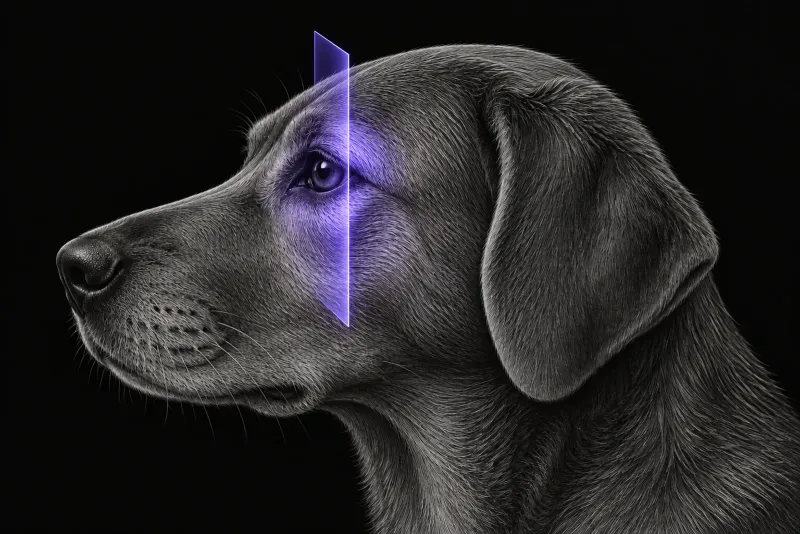
Neoplasia and inflammation are both common
Exophthalmos in a dog can mean a tumour or an infection, and the two are close in frequency:
- In 81 referral dogs with orbital disease that had CT or MRI (Coall et al., Veterinary Ophthalmology, 2022), inflammatory disease was more frequent than neoplasia (59% vs 41%). Orbital cellulitis was the most common diagnosis (36 of 81, 44.4%). The most common tumours were sarcoma (10 of 30) and carcinoma (7 of 30).
- In 66 dogs with retrobulbar disease (Winer et al., Frontiers in Veterinary Science, 2018), 41 had disease that started in the retrobulbar space: 19 neoplastic, 19 infectious or inflammatory and 3 traumatic. Of the 25 dogs with secondary retrobulbar disease, 21 had neoplasia.
- Clinical history helps. In a prospective study of 45 dogs with unilateral orbital disease (Boroffka et al., Journal of the American Veterinary Medical Association, 2007), dogs with neoplasia were older, had signs for longer and had a more progressive onset than dogs with non-neoplastic disease.
What CT shows: findings that shift the probability
| Finding on CT | Points towards |
|---|---|
| Orbital osteolysis | Neoplasia (Coall et al.; Winer et al.) |
| Orbital periosteal reaction or new bone | Neoplasia (Winer et al.) |
| A discrete retrobulbar mass | Neoplasia (Winer et al.) |
| Clearly delineated lesion margins | Neoplasia (odds ratio 45 on CT) (Boroffka et al.) |
| Intracranial extension | Neoplasia (Coall et al.) |
| Zygomatic salivary gland enlargement | Infection or inflammation (Winer et al.) |
| Retrobulbar mass effect | Infection or inflammation (Winer et al.) |
| Mandibular lymphadenopathy | Infection or inflammation (Winer et al.) |
CT findings overlap between retrobulbar diseases, so none of these signs is diagnostic on its own. CT showed extraorbital involvement more often than ultrasound, although the overall diagnostic value of the two was similar in that study.
Two specific patterns are worth knowing:
- Retrobulbar meningioma. In 15 dogs (Sidhu and DeJesus, Veterinary Radiology & Ultrasound, 2023), meningiomas were usually well-defined conical to ovoid masses along the optic nerve, expanding the extraocular muscle cone. Partial mineralisation was best seen on CT (9 dogs). Osteolysis was rare (3 dogs), yet extension into the cranial cavity was found in 6 dogs, including some without osteolysis.
- Bilateral zygomatic sialadenitis. In 20 dogs (Enache et al., Journal of Small Animal Practice, 2025), the most common signs were pain on opening the mouth (18 of 20) and exophthalmos (15 of 20). On CT, the zygomatic glands were all hyperattenuating and contrast enhancing. Fifteen of 20 dogs had at least one concurrent systemic disease.
Where MRI adds detail
MRI shows the soft tissue extent of inflammation well. In 31 dogs with retrobulbar inflammation on low-field MRI (Fischer et al., Journal of Small Animal Practice, 2019), extraocular myositis was present in 27. Of 25 dogs with exophthalmos, 14 had optic nerve swelling, and five of these lost vision permanently. No dog without nerve swelling lost vision.
Suspected foreign bodies are often not confirmed. MRI overestimated their presence: a foreign body was found at surgery in only one dog. In the Australian series, a foreign body was suspected in 36.1% of dogs with cellulitis, but only three were retrieved at orbitotomy.
Because meningiomas can extend into the cranial cavity without bone lysis, MRI is worth discussing when CT suggests a mass along the optic nerve.
Orbit CT protocol at a glance
Orbital CT covers more than the orbit, because several findings that shift the probability lie beyond it: the cranial cavity for intracranial extension, and the zygomatic salivary glands and mandibular lymph nodes for signs of inflammation. Contrast enhancement is part of the picture, as in zygomatic sialadenitis. CT can also plan the route for a core needle biopsy while the dog is still on the table.
What the radiologist looks at
The findings are weighed together, because no single sign is diagnostic.
- Bone. Orbital osteolysis, periosteal reaction or new bone, which point towards neoplasia.
- The lesion itself. A discrete mass and clearly delineated margins favour neoplasia, while a retrobulbar mass effect favours infection or inflammation.
- The optic nerve. A conical or ovoid mass along it, with partial mineralisation, fits retrobulbar meningioma.
- Beyond the orbit. Intracranial extension, zygomatic salivary gland change and mandibular lymph nodes, plus any other extraorbital involvement.
What your report will include
Every Veheri case is read by a radiologist or neurologist, and when you order you can choose Diplomate-only reporting or the full specialist panel. You can chat directly with the specialist assigned to your case, and a simplified pet owner report helps you explain the findings to the owner.
CT narrows the differential and plans the route to the lesion. It does not give a histological diagnosis. When you take the report forward:
- Biopsy. In 23 canine orbital masses (Cirla et al., Open Veterinary Journal, 2016), ultrasound-guided core needle biopsy was performed with the dog still on the CT table. CT was used to choose a trans-orbital, trans-conjunctival or trans-masseteric approach. Every sample was sufficient for histology and matched the excisional biopsy.
- Culture. When orbital cellulitis is suspected, the Australian authors recommend bacterial culture and susceptibility testing. Multidrug-resistant bacteria were found in three samples and changed treatment.
- MRI. Worth discussing when a mass lies along the optic nerve, since meningiomas can reach the cranial cavity without bone lysis.
- Bilateral zygomatic sialadenitis. Look for concurrent systemic disease, present in 15 of 20 dogs.
- Talking to the owner: in orbital inflammation, permanent vision loss occurred only in dogs with optic nerve swelling.
What to include when you request an orbital CT report
- signalment: age, breed and sex
- duration and onset of signs: sudden or progressive
- pain on opening the mouth, third eyelid protrusion and resistance to retropulsion
- vision in both eyes
- oral and dental findings, and any nasal signs
- concurrent systemic disease
- previous treatment and response, including antibiotics
How Veheri can help
Veheri’s radiologists report veterinary CT studies remotely for clinics, and can support your team with CT scanning protocols for head CT. For related reading, see MRI of the canine orbit and when a CT scan is indicated in veterinary medicine.
Sources
Each claim in this guide links to its source. The full citations:
- Coall SM, Premont JE, Flatz K, et al. Clinical, advanced imaging data and outcome of inflammatory and neoplastic orbital disease in 81 dogs and 16 cats in Australia (2010-2019). Vet Ophthalmol. 2022;25 Suppl 1:37-50. doi:10.1111/vop.12936
- Winer JN, Verstraete FJM, Cissell DD, et al. Clinical features and computed tomography findings are utilized to characterize retrobulbar disease in dogs. Front Vet Sci. 2018;5:186. doi:10.3389/fvets.2018.00186
- Boroffka SA, Verbruggen AM, Grinwis GC, et al. Assessment of ultrasonography and computed tomography for the evaluation of unilateral orbital disease in dogs. J Am Vet Med Assoc. 2007;230(5):671-80. doi:10.2460/javma.230.5.671
- Sidhu A, DeJesus AA. Computed tomographic, magnetic resonance imaging, and ultrasonographic characteristics of retrobulbar meningiomas in 15 dogs. Vet Radiol Ultrasound. 2023;64(6):1044-1054. doi:10.1111/vru.13311
- Enache AE, Maini S, Pivetta M, et al. Canine bilateral zygomatic sialadenitis: 20 cases (2000-2019). J Small Anim Pract. 2025;66(6):396-411. doi:10.1111/jsap.13844
- Fischer MC, Busse C, Adrian AM. Magnetic resonance imaging findings in dogs with orbital inflammation. J Small Anim Pract. 2019;60(2):107-115. doi:10.1111/jsap.12929
- Cirla A, Rondena M, Bertolini G. Automated tru-cut imaging-guided core needle biopsy of canine orbital neoplasia. A prospective feasibility study. Open Vet J. 2016;6(2):114-20. doi:10.4314/ovj.v6i2.7
Related reading
More imaging guides
All imaging guides
MRI of the canine orbit and optic nerve, compared with CT
When MRI or CT best answers canine orbital and optic nerve questions, which signs point to tumour or inflammation, and when sampling is still needed.

CT of adrenal masses in dogs
What contrast CT shows in a dog with an adrenal mass, from caval invasion and adhesions to tumour type, and where its accuracy runs out before adrenalectomy.

Brain CT in dogs when MRI is not available
What brain CT can and cannot detect in dogs when MRI is not available, for masses, stroke, inflammation and head trauma, and when MRI or CSF is still needed.