
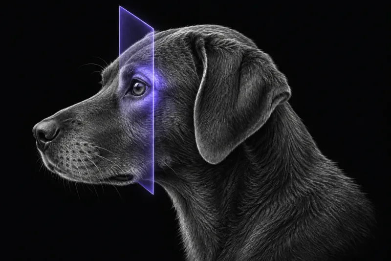
What is behind orbital signs in dogs
The mix of diagnoses depends on who is referred and how they are imaged.
- In 81 dogs and 16 cats with orbital disease that had CT (84 animals) or MRI (13) (Coall et al., Veterinary Ophthalmology), inflammatory disease was more common than neoplasia in dogs (59% versus 41%). Orbital cellulitis was the most common diagnosis in dogs (36 of 81, 44.4%). The most common canine tumours were sarcoma (10 of 30) and carcinoma (7 of 30).
- In 79 dogs and 13 cats that had MRI after referral to an ophthalmologist (Armour et al., Veterinary Ophthalmology, 2011), neoplasia was diagnosed in 53 of 92 (57.6%), suspected optic neuritis in 13 (14.1%) and orbital cellulitis in 12 (13.0%).
Both are large enough to show that inflammation, neoplasia and optic nerve disease all need to be on the list when a dog presents with orbital signs.
MRI or CT: match the scan to the question
We did not find a study that compares MRI and CT head to head in the canine orbit. The choice rests on what each modality has shown on its own.
| Clinical question | Evidence |
|---|---|
| Bone lysis, periosteal reaction, mineralisation | CT findings of orbital osteolysis, periosteal reaction and a retrobulbar mass were significantly associated with neoplasia in 66 dogs (Winer et al., Frontiers in Veterinary Science, 2018). In retrobulbar meningiomas (Sidhu and DeJesus, Veterinary Radiology & Ultrasound, 2023), mineralisation was best seen on CT in nine dogs and only suspected from susceptibility artefact on MRI in three. |
| Optic nerve and chiasm | In 18 dogs with presumed optic neuritis (Muñiz Moris et al., Frontiers in Veterinary Science, 2021), low-field MRI showed contrast enhancement of both optic nerves in 11, chiasmal enhancement in 6, changes in the fluid around the nerve in 10 and concurrent brain lesions in 13. In 28 dogs, 48 nerves (Bedos et al., Journal of Small Animal Practice, 2020), MRI showed nerve enlargement in 32 nerves (67%) and enhancement in 28 (58%). |
| Spread into the cranial cavity | In four dogs with chronic orbital inflammation (Kneissl et al., Veterinary Radiology & Ultrasound, 2007), MRI showed extension through the orbital fissure in all four. Six of 15 retrobulbar meningiomas extended into the cranial cavity, sometimes without osteolysis. |
| Blindness with no other deficits | In seven dogs and cats with acute postretinal blindness (Seruca et al., Veterinary Ophthalmology, 2010), MRI showed visual pathway lesions in six: five consistent with intracranial tumours and one with bilateral optic neuritis. |
In short: CT is strong for bone and mineral, and MRI is usually preferred when the optic nerve, chiasm or brain may be involved.
Orbital MRI protocol at a glance
Orbital MRI has to show the orbit, the optic pathway and the cranial cavity in one study. Extension through the orbital fissure was best identified on transverse T2-weighted, dorsal STIR or post-contrast images. Because concurrent brain lesions were common in optic neuritis, an orbital MRI for vision loss usually includes the brain.
What the radiologist looks at
- Osteolysis and intracranial extension. Both were significantly associated with neoplasia in dogs.
- Margins. Well-defined margins were the most discriminating feature on both ultrasound and CT in 45 dogs with unilateral orbital disease (Boroffka et al., Journal of the American Veterinary Medical Association, 2007), with neoplastic lesions more often clearly delineated. CT more often showed extraorbital involvement.
- Inflammatory patterns. On CT these included zygomatic salivary gland enlargement, retrobulbar mass effect and mandibular lymphadenopathy.
- Tumour type. In the 92-patient MRI review:
- carcinomas and sarcomas showed bony lysis and intracranial or sinonasal extension
- lymphoma was generally unilateral, less invasive and arose from the ventromedial orbit
- presumptive meningiomas often showed a dural tail sign
- Meningioma. Retrobulbar meningiomas were usually well-defined conical to ovoid masses along the optic nerve that expanded the muscle cone, with moderate to marked enhancement on MRI in all cases.
These findings overlap between diseases, and the CT study authors say so plainly.
What your report will include
Every Veheri case is read by a radiologist or neurologist, and when you order you can choose Diplomate-only reporting or the full specialist panel. Neurology reporting covers CT and MRI. You can chat directly with the specialist assigned to your case, and a simplified pet owner report helps you explain the findings to the owner.
When you take the report forward:
- Sample suspected cellulitis. A foreign body was suspected in 36.1% of dogs with orbital cellulitis, yet only 3 were retrieved at orbitotomy. Multidrug-resistant bacteria changed treatment in 3 samples, and the authors recommend culture and susceptibility testing when cellulitis is suspected.
- Optic neuritis remains a presumptive diagnosis. MRI changes can be subtle.
- CSF without pleocytosis does not exclude optic neuritis. In the 28-dog series, cerebrospinal fluid showed pleocytosis in only 11 of 25 dogs sampled.
What to include when you request an orbital MRI report
- signalment: age, breed and sex
- laterality, exophthalmos or enophthalmos, third eyelid protrusion and pain on opening the mouth
- onset and duration: dogs with neoplasia were older and had a longer, more progressive history
- vision, pupillary light reflexes and fundic findings
- nasal or oral signs, since dogs with retrobulbar disease showed combinations of ocular, oral and nasal signs
- any neurological signs, and CSF or infectious disease results
- a previous CT, if one was done
How Veheri can help
Veheri provides veterinary teleradiology and teleneurology and reports MRI and CT studies of the head remotely for clinics. If CT is your first scan, see CT of the canine orbit, and for vision loss with other neurological signs, when veterinary MRI is the right move.
Sources
Each claim in this guide links to its source. The full citations:
- Coall SM, Premont JE, Flatz K, et al. Clinical, advanced imaging data and outcome of inflammatory and neoplastic orbital disease in 81 dogs and 16 cats in Australia (2010-2019). Vet Ophthalmol. 25 Suppl 1:37-50 (published online 2021). doi:10.1111/vop.12936
- Armour MD, Broome M, Dell'Anna G, et al. A review of orbital and intracranial magnetic resonance imaging in 79 canine and 13 feline patients (2004-2010). Vet Ophthalmol. 2011;14(4):215-26. doi:10.1111/j.1463-5224.2010.00865.x
- Winer JN, Verstraete FJM, Cissell DD, et al. Clinical features and computed tomography findings are utilized to characterize retrobulbar disease in dogs. Front Vet Sci. 2018;5:186. doi:10.3389/fvets.2018.00186
- Sidhu A, DeJesus AA. Computed tomographic, magnetic resonance imaging, and ultrasonographic characteristics of retrobulbar meningiomas in 15 dogs. Vet Radiol Ultrasound. 2023;64(6):1044-1054. doi:10.1111/vru.13311
- Muñiz Moris L, Cherubini GB, Caine A. Low-field magnetic resonance imaging findings in 18 dogs with presumed optic neuritis. Front Vet Sci. 2021;7:585828. doi:10.3389/fvets.2020.585828
- Bedos L, Tetas R, Crespo V, Shea A. Presumed optic neuritis of non-infectious origin in dogs treated with immunosuppressive medication: 28 dogs (2000-2015). J Small Anim Pract. 2020;61(11):676-683. doi:10.1111/jsap.13233
- Kneissl S, Konar M, Fuchs-Baumgartinger A, Nell B. Magnetic resonance imaging features of orbital inflammation with intracranial extension in four dogs. Vet Radiol Ultrasound. 2007;48(5):403-8. doi:10.1111/j.1740-8261.2007.00267.x
- Seruca C, Ródenas S, Leiva M, et al. Acute postretinal blindness: ophthalmologic, neurologic, and magnetic resonance imaging findings in dogs and cats (seven cases). Vet Ophthalmol. 2010;13(5):307-14. doi:10.1111/j.1463-5224.2010.00814.x
- Boroffka SA, Verbruggen AM, Grinwis GC, et al. Assessment of ultrasonography and computed tomography for the evaluation of unilateral orbital disease in dogs. J Am Vet Med Assoc. 2007;230(5):671-80. doi:10.2460/javma.230.5.671
Related reading
More imaging guides
All imaging guides
CT of the canine orbit for exophthalmos and retrobulbar disease
How CT helps separate orbital neoplasia from cellulitis and other inflammation in dogs with exophthalmos, where MRI adds detail, and how CT guides sampling.
MRI of the canine brachial plexus for peripheral nerve sheath tumours
How MRI finds peripheral nerve sheath tumours of the canine brachial plexus, what the radiologist looks for, and what imaging can say about grade and extent.
Brain MRI for dogs with seizures
Which dogs with seizures need brain MRI, what the evidence says about diagnostic yield, what the radiologist looks for and what your report will include.