
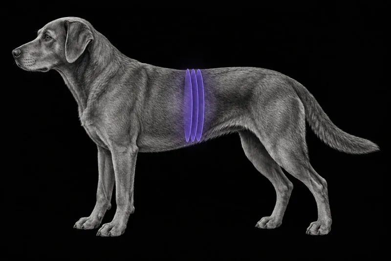
Why CT angiography for a suspected shunt
Ultrasound is often the first imaging test when a portosystemic shunt is suspected. It misses a meaningful share of shunts, though, and a surgeon usually needs the exact anatomy, not only a yes or no.
In 76 dogs investigated for a suspected shunt (Kim et al., Veterinary Radiology & Ultrasound, 2013), CT angiography (CTA) had a sensitivity of 96% against 68% for abdominal ultrasound. Specificity was similar (89% and 84%). CTA was 5.5 times more likely than ultrasound to correctly establish whether a shunt was present.
| Question | CTA | Ultrasound |
|---|---|---|
| Correct shunt origin (congenital shunts) | 15 of 16 dogs | 24 of 30 dogs |
| Correct shunt insertion (congenital shunts) | 15 of 16 dogs | 20 of 33 dogs |
| Multiple acquired shunts seen | 4 of 5 dogs | 1 of 6 dogs |
Source: Kim et al.
Is the extra anaesthetic safe? In 124 dogs treated for a single congenital extrahepatic shunt (Brunson et al., Canadian Veterinary Journal, 2016), major postoperative complications occurred in 4.7% of dogs that had preoperative CTA and 9.9% of those that did not, a difference that was not significant. Surgical time was similar (84 and 81 minutes). The authors concluded that anaesthetised preoperative CTA appears safe and does not affect surgical time, complication rate or outcome.
Classifying extrahepatic shunts
-
In 25 dogs (Nelson and Nelson, Veterinary Radiology & Ultrasound, 2011), CTA identified six general extrahepatic shunt types. Two types had large anastomosing loops that would matter at surgery, and in 2 dogs no portal vein could be identified cranial to the shunt origin.
-
In a multi-institutional series of 1082 dogs (Weisse et al., Veterinary Radiology & Ultrasound, 2024), 45 different extrahepatic anatomies were found. Five classes made up 85% of shunts:
- left gastric-phrenic (27%)
- left gastric-azygos (19%)
- left gastric-caval (15%)
- aberrant left gastric-caval with right gastric vein (12%)
- the same with an added short gastric vein (11%)
The left gastric vein was involved in the origin in 95% of the described classifications. Shunt types differed by age, sex, breed, weight and portal perfusion score.
Classifying intrahepatic shunts
In 90 dogs with intrahepatic shunts on CTA (Plested et al., Veterinary Radiology & Ultrasound, 2020), 92% inserted into a primary hepatic vein or phrenic vein and 8% directly into the intrahepatic caudal vena cava. Right divisional shunts were most common (44%), then left (33%) and central (13%). Multiple insertion sites were seen in 9%. The authors suggest that naming the receiving hepatic vein makes classification more consistent and may influence how the shunt is attenuated.
Portal vasculature CT protocol at a glance
CTA shows normal portal vessels, congenital intrahepatic and extrahepatic shunts, arterioportal fistulae and multiple acquired shunts. According to a review of CT diagnosis of portosystemic shunts (Zwingenberger, Veterinary Clinics of North America: Small Animal Practice, 2009), contrast timing, the bolus and a respiratory pause are essential to a diagnostic study.
What the radiologist looks at
- Is there a shunt, and what kind? Whether a shunt is present, and whether it is single congenital or multiple acquired.
- Origin and insertion. The portal vessel of origin and the systemic vessel of insertion, using the extrahepatic classes from the 1082-dog series.
- For intrahepatic shunts, the division and the receiving hepatic vein.
- What the surgeon will meet. Loops and additional contributing vessels, which would matter at surgery.
- The intrahepatic portal branches. In 146 dogs with an extrahepatic shunt (Takeuchi et al., American Journal of Veterinary Research, 2025), the CTA grade of intrahepatic portal branching correlated with portal pressure during temporary occlusion and differed between dogs that had partial and complete ligation.
- CTA against portovenography. In 49 dogs and 10 cats with a single extrahepatic shunt (Parry and White, Journal of Small Animal Practice, 2017), CTA showed all four portal vein tributaries. It outperformed intraoperative mesenteric portovenography for the intrahepatic portal vein, except in shunts involving the right gastric vein.
What your report will include
Every Veheri case is read by a radiologist or neurologist, and when you order you can choose Diplomate-only reporting or the full specialist panel. You can chat directly with the specialist assigned to your case, and a simplified pet owner report helps you explain the findings to the owner.
When you take the report forward:
- Absent portal branches on CTA do not on their own rule out attenuation. In 7 dogs with a single extrahepatic shunt and absent portal branches (Bae et al., American Journal of Veterinary Research, 2025), 6 had successful attenuation, and in 2 dogs portovenography showed intrahepatic portal vessels only after temporary shunt occlusion. The authors support intraoperative evaluation such as portovenography to guide the decision.
- Portovenography at surgery still adds value: after temporary full ligation it gives useful information.
- Branching grade is not a prognosis. It did not predict complications or prognosis.
What to include when you request a CTA report
- signalment: age, breed and sex
- clinical signs
- bile acids, ammonia and other blood results
- previous ultrasound findings
- any previous shunt surgery and the device used
- whether surgical or interventional attenuation is planned
How Veheri can help
Veheri’s radiologists report veterinary CT studies remotely for clinics, and we offer expert support for CT protocols, positioning and acquisition. For the feline version, see CT of the portal vasculature in cats, and for abdominal scanning, see optimising routine abdominal CT protocols.
Sources
Each claim in this guide links to its source. The full citations:
- Kim SE, Giglio RF, Reese DJ, et al. Comparison of computed tomographic angiography and ultrasonography for the detection and characterization of portosystemic shunts in dogs. Vet Radiol Ultrasound. 2013;54(6):569-74. doi:10.1111/vru.12059
- Zwingenberger A. CT diagnosis of portosystemic shunts. Vet Clin North Am Small Anim Pract. 2009;39(4):783-92. doi:10.1016/j.cvsm.2009.04.008
- Brunson BW, Case JB, Ellison GW, et al. Evaluation of surgical outcome, complications, and mortality in dogs undergoing preoperative computed tomography angiography for diagnosis of an extrahepatic portosystemic shunt: 124 cases (2005-2014). Can Vet J. 2016;57(1):59-64. PMID: 26740699. https://pubmed.ncbi.nlm.nih.gov/26740699/
- Nelson NC, Nelson LL. Anatomy of extrahepatic portosystemic shunts in dogs as determined by computed tomography angiography. Vet Radiol Ultrasound. 2011;52(5):498-506. doi:10.1111/j.1740-8261.2011.01827.x
- Weisse C, Fox-Alvarez WA, Grosso FRV, et al. Anatomical classification of canine congenital extrahepatic portosystemic shunts based on CT angiography: a SVSTS and VIRIES multi-institutional study in 1082 dogs. Vet Radiol Ultrasound. 2024;65(6):702-712. doi:10.1111/vru.13415
- Plested MJ, Zwingenberger AL, Brockman DJ, et al. Canine intrahepatic portosystemic shunt insertion into the systemic circulation is commonly through primary hepatic veins as assessed with CT angiography. Vet Radiol Ultrasound. 2020;61(5):519-530. doi:10.1111/vru.12892
- Takeuchi R, Ishigaki K, Iida K, et al. Use of computed tomography angiography has potential in planning surgical treatment of an extrahepatic portosystemic shunt in dogs. Am J Vet Res. 2025;86(4). doi:10.2460/ajvr.24.11.0333
- Parry AT, White RN. Comparison of computed tomographic angiography and intraoperative mesenteric portovenography for extrahepatic portosystemic shunts. J Small Anim Pract. 2017;58(1):49-55. doi:10.1111/jsap.12596
- Bae S, Culp WTN, Zwingenberger AL. Dogs with congenital extrahepatic shunts and absent portal vasculature noted on computed tomographic angiography can successfully undergo surgical attenuation. Am J Vet Res. 2025;86(11). doi:10.2460/ajvr.25.04.0146
Related reading
More imaging guides
All imaging guides
CT angiography of the feline portal vasculature for portosystemic shunts
How CT angiography maps portosystemic shunts in cats, how it compares with ultrasound and portovenography, and which feline anatomy the report must cover.

CT of adrenal masses in dogs
What contrast CT shows in a dog with an adrenal mass, from caval invasion and adhesions to tumour type, and where its accuracy runs out before adrenalectomy.

Brain CT in dogs when MRI is not available
What brain CT can and cannot detect in dogs when MRI is not available, for masses, stroke, inflammation and head trauma, and when MRI or CSF is still needed.