
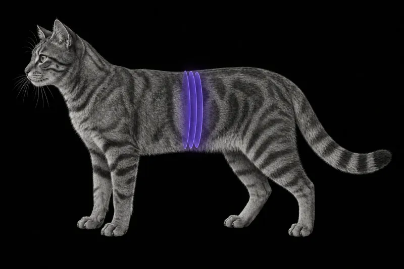
Why feline shunt anatomy needs its own map
Shunts in cats are not small versions of shunts in dogs. In a multi-institutional study of 231 cats with congenital extrahepatic portosystemic shunts on CT angiography (Weisse et al., Veterinary Radiology & Ultrasound, 2024), 25 different shunt anatomies were found. Five classes accounted for 78% of shunts, and the most common class alone for 53%. The left gastric vein was involved in the shunt origin in 75% of the described classifications. The five most common types differed in age at CT, breed and subjective portal perfusion score.
Normal feline anatomy also varies. In 52 cats without vascular anomalies (Dekkers et al., Journal of Feline Medicine and Surgery, 2025):
- the left gastric vein entered the extrahepatic portal vein directly in 42 cats
- in 8 cats a single left gastric vein entered the splenic vein, as is typical in dogs
- the gastroduodenal vein entered the portal vein in 34 cats; in the other 18, the right gastric and cranial pancreaticoduodenal veins entered together
A report that names the vessel of origin and the vessel of insertion uses this feline anatomy, not the canine template.
When to image
Blood tests raise the suspicion; imaging confirms and maps. In 85 cats tested for a shunt (Ruland et al., Veterinary Clinical Pathology), serum bile acids at the laboratory upper reference limit had 100% sensitivity and 71% specificity for a shunt, and fasting ammonia 83% sensitivity and 76% specificity. With optimised cut-offs, bile acids reached 100% sensitivity and 84% specificity in cats.
Ultrasound, CTA and portovenography
| Method | What the evidence shows |
|---|---|
| Ultrasound | In 85 dogs and 17 cats with a suspected shunt (d’Anjou et al., Veterinary Radiology & Ultrasound, 2004), 92% sensitive, 98% specific, 95% accurate. When ultrasound found a shunt, extrahepatic, intrahepatic and multiple acquired shunts were correctly told apart in 53 of 54 patients. Cats were a small part of this study. |
| CT angiography | Showed all four portal vein tributaries and sub-tributaries in 49 dogs and 10 cats with single extrahepatic shunts (Parry and White, Journal of Small Animal Practice). |
| Intraoperative mesenteric portovenography | In the same 10 cats, did not show the gastroduodenal vein in any cat, and showed the splenic vein in 8 and the caudal mesenteric vein in 2. |
Not every shunt is congenital
Splenosystemic shunts in cats (Palerme et al., Journal of Veterinary Internal Medicine, 2013) were identified in 1.3% of cats having abdominal ultrasound in one hospital. Older, spayed female cats were over-represented, and 42% had a hepatopathy with the potential for portal hypertension. Their signalment and anatomy differed from those of cats with single congenital shunts.
Portal CT angiography protocol at a glance
Shunt mapping in cats relies on CT angiography of the portal system, which showed all four portal vein tributaries and sub-tributaries in cats with single extrahepatic shunts. When diagnostic CTA has been performed before surgery, the same authors conclude that diagnostic portovenography before ligation is not indicated.
What the radiologist looks at
- Origin and insertion of the shunt, named with feline anatomy, including whether the left gastric vein is involved.
- Contributing portal vessels, which vary even in normal cats.
- Portal vein size. In 114 cats (Jeong et al., Journal of Veterinary Science, 2024), normal cats had a portal vein diameter of 5.23 ± 0.77 mm and a portal vein to aorta (PV/Ao) ratio of 1.46 ± 0.19. Between normal cats and cats with symptomatic shunts, a cut-off of 4.115 mm for portal vein diameter had 100% sensitivity and 97.7% specificity. A PV/Ao cut-off of 1.170 had 90% sensitivity and 92.1% specificity. These indices differed significantly between dogs and cats, so canine reference values should not be assumed to apply to cats.
- Shunt type and the kidneys. In 83 cats with a single extrahepatic shunt (Kim et al., Journal of Veterinary Medical Science, 2026), the left gastrocaval group had the lowest median PV/Ao ratio (0.56) and renomegaly in all cats. The splenosystemic group had the highest ratio (1.12) and renomegaly in 12.8%. Kidney size and portal perfusion were inversely correlated.
- Portal vein patency. In two cats with splenosystemic shunts and hepatic encephalopathy (Sakamoto et al., Journal of Small Animal Practice, 2022), CTA showed thrombi as non-enhancing structures inside the main portal vein.
What your report will include
Every Veheri case is read by a radiologist or neurologist, and when you order you can choose Diplomate-only reporting or the full specialist panel. You can chat directly with the specialist assigned to your case, and a simplified pet owner report helps you explain the findings to the owner.
When you take the report forward:
- At surgery, portovenography after temporary full ligation still gives useful information.
- A portal vein thrombus can be followed: follow-up CTA tracked the course of the thrombi.
- CTA shows anatomy. It does not replace the clinical and laboratory assessment of the cat, and the portal perfusion score in the classification study was subjective.
What to include when you request a CTA report
- signalment: age, breed and sex
- neurological or gastrointestinal signs
- bile acids and ammonia results
- ultrasound findings, including any suspected shunt vessel
- previous shunt surgery or attenuation devices
- the question for the surgeon: vessel of origin, vessel of insertion, contributing portal vessels, portal vein size and patency
How Veheri can help
Veheri’s radiologists report veterinary CT studies remotely for clinics, and offer expert support with CT protocols, positioning and acquisition. For dogs, see CT angiography of the canine portal vasculature, and for abdominal scanning, our abdominal CT protocols article.
Sources
Each claim in this guide links to its source. The full citations:
- Weisse C, Asano K, Ishigaki K, et al. Anatomical classification of feline congenital extrahepatic portosystemic shunts based on CT angiography: a SVSTS and VIRIES multi-institutional study in 231 cats. Vet Radiol Ultrasound. 2024;65(4):359-368. doi:10.1111/vru.13363
- Dekkers MM, Devriendt N, de Rooster H, et al. Anatomical variations in the tributaries of the normal feline extrahepatic portal system: evaluation with CT angiography. J Feline Med Surg. 2025;27(9):1098612X251368447. doi:10.1177/1098612X251368447
- Ruland K, Fischer A, Hartmann K. Sensitivity and specificity of fasting ammonia and serum bile acids in the diagnosis of portosystemic shunts in dogs and cats. Vet Clin Pathol. 39(1):57-64 (published online 2009). doi:10.1111/j.1939-165X.2009.00178.x
- d'Anjou MA, Penninck D, Cornejo L, Pibarot P. Ultrasonographic diagnosis of portosystemic shunting in dogs and cats. Vet Radiol Ultrasound. 2004;45(5):424-37. doi:10.1111/j.1740-8261.2004.04076.x
- Parry AT, White RN. Comparison of computed tomographic angiography and intraoperative mesenteric portovenography for extrahepatic portosystemic shunts. J Small Anim Pract. 58(1):49-55 (published online 2016). doi:10.1111/jsap.12596
- Jeong E, Chung JY, Ahn JO, et al. Computed tomographic evaluation of portal vein indices in cats with the extrahepatic portosystemic shunts. J Vet Sci. 2024;25(3):e37. doi:10.4142/jvs.24038
- Kim M, Noh D, Choi S, et al. Renomegaly severity and prevalence differ by shunt type and inversely correlate with CT-assessed portal perfusion in cats with extrahepatic portosystemic shunt. J Vet Med Sci. 2026;88(3):432-438. doi:10.1292/jvms.25-0495
- Palerme JS, Brown JC, Marks SL, Birkenheuer AJ. Splenosystemic shunts in cats: a retrospective of 33 cases (2004-2011). J Vet Intern Med. 2013;27(6):1347-53. doi:10.1111/jvim.12188
- Sakamoto Y, Ishikawa C, Nakayama T, Sakai M. Computed tomographic features of portal vein thrombosis in two cats with splenosystemic shunts. J Small Anim Pract. 2022;63(7):563-568. doi:10.1111/jsap.13470
Related reading
More imaging guides
All imaging guides
CT angiography of the canine portal vasculature for portosystemic shunts
How CT angiography compares with ultrasound for congenital portosystemic shunts in dogs, how shunts are classified, and what the surgeon needs from the report.
CT of the feline interscapular region for injection-site sarcoma
What contrast CT shows in feline injection-site sarcoma, how it changes surgical and radiotherapy planning, and where imaging cannot replace histology.
Thoracic CT in cats for lung tumours, airway and interstitial disease
What thoracic CT adds over radiographs in cats with lung tumours, bronchial or interstitial disease, how anaesthesia affects the lungs, and where CT stops.