
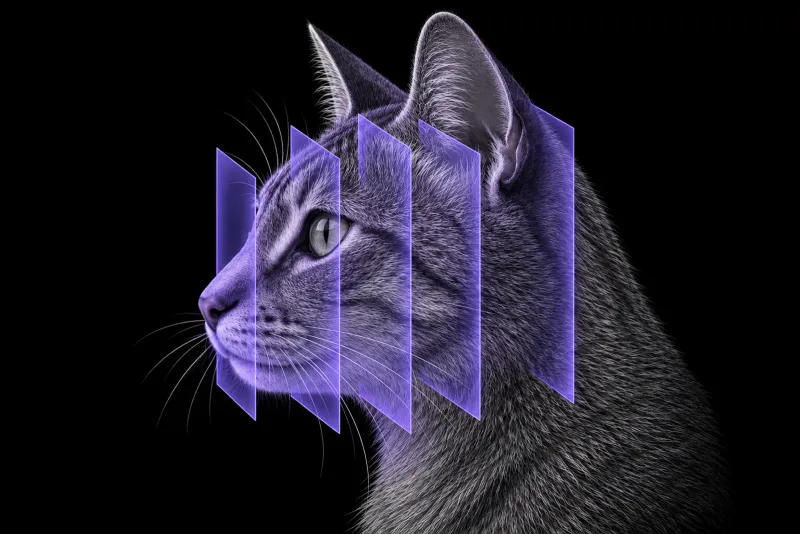
Why feline head trauma is a CT question
Skull fractures in cats are usually multiple. In 75 cats with CT-confirmed skull fractures (Knight and Meeson, Journal of Feline Medicine and Surgery), 89% had fractures in multiple bones. The mandible, the upper jaw (maxilla, incisive and nasal bones) and the craniofacial region were most often affected, and 56% had a temporomandibular joint (TMJ) injury. Road traffic accidents were the cause in 89% of cats and were associated with concurrent ophthalmic, neurological and thoracic injuries.
Cats also break their skulls differently from dogs. In 95 cats and 91 dogs with traumatic skull fractures on CT (Amengual-Batle et al., Journal of Veterinary Internal Medicine, 2020), the face and skull base were more often affected in cats, and cats more often had multiple fractures.
Radiography compared with CT
A review for general practitioners (Rizkallal and Lafuente, Journal of Feline Medicine and Surgery, 2020) notes that radiography may help, but some injuries are hard to see because soft tissue and bone superimpose. CT is a more sensitive technique for skull injuries. A cadaver study (Hecht et al., Frontiers in Veterinary Science, 2021) describes CT as the imaging modality of choice for acute head trauma. MRI agreed with CT on the presence or absence of a fracture in 93.5% of assessments overall, but only 87.6% on post-trauma studies.
The same review stresses that the cat must first be stabilised, with assessment of the respiratory, cardiovascular, neurological and ophthalmic systems. Imaging follows once the cat is stable enough for anaesthesia.
Fracture patterns CT should map
| Region | What the evidence shows |
|---|---|
| Mandible | Fractured in 40 of 48 cats (Weichert et al., Veterinary and Comparative Orthopaedics and Traumatology). In 109 cats with mandibular trauma (Evenhuis et al., Frontiers in Veterinary Science, 2025), the symphysis (55.0%), condylar process (49.5%) and mid ramus (48.6%) were most often injured. |
| TMJ | In 79 cats, 158 joints (Mestrinho et al., Journal of Feline Medicine and Surgery, 2022), injuries were unilateral in 70.9%. The condyle was fractured in 55.7% of joints, and 84.0% of these were intra-articular, with the medial half over-represented. Luxations occurred in 32.9% of cats, 87.0% of them rostrodorsal. |
| Temporal bone | Fractured in 30.4% of the 79 cats, and associated with road traffic accidents. |
| Orbit, nasopharynx, zygomatic arch | Fractured in 30, 29 and 12 of 48 cats. |
| Cranial vault | Associated with neurological signs, especially when depressed, and with death in the first week when fragmented or displaced (Amengual-Batle et al., 2020). |
In 36 of 48 cats, the fractures involved two or more functional systems, such as the oral cavity with the nasal airways, or the nasal airways with the orbit and eye. Bilateral TMJ injuries were 13.1 times more likely after high-rise trauma.
The brain
In 14 cats with traumatic brain injury that had head CT within 72 hours of admission (Mann et al., Journal of Feline Medicine and Surgery), findings included:
- lateral ventricle asymmetry or midline shift (42.8%)
- intracranial haemorrhage (35.7%)
- cranial vault fractures (14.2%), all depressed
Intracranial haemorrhage was negatively associated with short- and long-term survival.
Head CT protocol at a glance
CT is the modality of choice for acute head trauma, and it is done once the cat is stable enough for anaesthesia. Because most cats have fractures in several bones, the study should cover the whole skull, including both TMJs and the brain.
What the radiologist looks at
- Displacement. More severe displacement before surgery was associated with poor healing in the mid ramus and coronoid process.
- Palate and pterygoid region. Fractures of the palatine and pterygoid bones and hard palate injuries increased the risk of implant loosening and malocclusion. Internal upper jaw fractures and older age were risk factors for complications.
- The TMJ in detail. Whether a condylar fracture is intra-articular, and the direction of any luxation.
- The cranial vault and brain. Depressed, fragmented or displaced vault fractures and intracranial haemorrhage are linked to neurological signs and survival.
What your report will include
Every Veheri case is read by a radiologist or neurologist, and when you order you can choose Diplomate-only reporting or the full specialist panel. Neurology reporting covers CT and MRI. You can chat directly with the specialist assigned to your case, and a simplified pet owner report helps you explain the findings to the owner.
When you take the report forward:
- Mandibular fractures. Open reduction and internal fixation gave adequate healing more often than maxillomandibular fixation, and persistent malocclusion was less common (9.1% versus 53.9%).
- TMJ luxation. In a study of cats with TMJ luxation (Jung et al., Frontiers in Veterinary Science, 2023), successful reduction was significantly associated with the time from injury to treatment, and concurrent dental and maxillofacial injuries were common.
- MRI may be chosen in selected cases.
- Talking to the owner: in the larger fracture series, 84.9% of the dogs and cats survived the first week, and the authors advise that a skull fracture alone should not be considered a negative prognostic factor. Keep in mind that the brain injury data come from 14 cats, and most of the feline fracture series cited here are retrospective.
What to include when you request a head CT report
- signalment: age, breed and sex
- cause of trauma: road traffic accident, fall from height or animal altercation
- time since injury
- neurological status and any modified Glasgow Coma Scale score
- occlusion and whether the cat can open and close its mouth
- ocular and thoracic findings
- whether surgical planning or a prognosis question is the priority
How Veheri can help
Veheri works with veterinary radiologists and neurologists and reports head CT studies remotely for clinics. For dogs, see CT of the canine temporomandibular joint and CT of the canine skull after head trauma.
Sources
Each claim in this guide links to its source. The full citations:
- Knight R, Meeson RL. Feline head trauma: a CT analysis of skull fractures and their management in 75 cats. J Feline Med Surg. 21(12):1120-1126 (published online 2018). doi:10.1177/1098612X18819183
- Amengual-Batle P, José-López R, Durand A, et al. Traumatic skull fractures in dogs and cats: a comparative analysis of neurological and computed tomographic features. J Vet Intern Med. 2020;34(5):1975-1985. doi:10.1111/jvim.15838
- Rizkallal C, Lafuente P. Feline skull injuries: treatment goals and recommended approaches. J Feline Med Surg. 2020;22(3):229-240. doi:10.1177/1098612X20903602
- Hecht S, Anderson KM, Castel A, et al. Agreement of magnetic resonance imaging with computed tomography in the assessment for acute skull fractures in a canine and feline cadaver model. Front Vet Sci. 2021;8:603775. doi:10.3389/fvets.2021.603775
- Weichert S, Kiefer I, Theyse LFH. Assessment of fracture distribution and involvement of functional systems following craniomaxillofacial trauma in 48 cats. Vet Comp Orthop Traumatol. 39(2):67-74 (published online 2025). doi:10.1055/a-2651-6219
- Evenhuis JV, Vincek A, Goldschmidt S, et al. Clinical and diagnostic imaging outcomes of mandibular fracture management in 109 cats. Front Vet Sci. 2025;12:1633636. doi:10.3389/fvets.2025.1633636
- Mestrinho LA, Sebastião SS, Zwierucho MA, et al. CT characterisation and classification of feline temporomandibular joint trauma: a case series of 79 cats. J Feline Med Surg. 2022;24(12):1181-1186. doi:10.1177/1098612X211066654
- Jung JW, Castejón-González AC, Stefanovski D, Reiter AM. Etiology, clinical presentation, and outcome of temporomandibular joint luxation in cats: 21 cases (2000-2018). Front Vet Sci. 2023;10:1274193. doi:10.3389/fvets.2023.1274193
- Mann O, Peery D, Bader Segev R, et al. CT findings and the prognostic value of the Koret CT score in cats with traumatic brain injury. J Feline Med Surg. 24(2):91-97 (published online 2021). doi:10.1177/1098612X211005306
Related reading
More imaging guides
All imaging guides
CT of the canine skull after head trauma
What head CT shows after trauma in dogs, which skull fracture patterns matter, and how far CT findings can support a prognosis alongside the neurological exam.
CT of the feline interscapular region for injection-site sarcoma
What contrast CT shows in feline injection-site sarcoma, how it changes surgical and radiotherapy planning, and where imaging cannot replace histology.
Thoracic CT in cats for lung tumours, airway and interstitial disease
What thoracic CT adds over radiographs in cats with lung tumours, bronchial or interstitial disease, how anaesthesia affects the lungs, and where CT stops.